
|
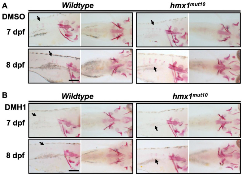
Figure 5 DMH1-mediated inhibition of BMP signaling reduces the progression of vertebral mineralization: Wildtype and hmx1mut10 embryos were treated with 50 μM DMH1 at 2 dpf and placed in E3 medium at 3 dpf. Alizarin red staining to detect mineralized structures was performed at 7 and 8 dpf (left, lateral view, right ventral view). Wildtype and hmx1mut10 control embryos treated with DMSO (A). Precocious vertebral mineralization was inhibited in DMH1-treated hmx1mut10 embryos at 7 dpf (B). At 8 dpf, treated hmx1mut10 embryos still presented a vertebral mineralization pattern similar to wildtype embryos (B). Bar, (A,B) 500 μm.