
- Title
-
MCRS1 associates with cytoplasmic dynein and mediates pericentrosomal material recruitment
- Authors
- Lee, S.H., Lee, M.S., Choi, T.I., Hong, H., Seo, J.Y., Kim, C.H., Kim, J.
- Source
- Full text @ Sci. Rep.
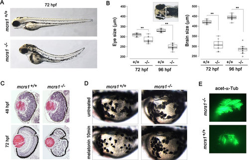
TALEN-mediated knockout of mcrs1 in zebrafish. (A) Homozygous mcrs1 mutant zebrafish exhibited a curved body axis at 72 hpf. (B) Measurement of the size of the eye and the brain (n>10 for each genotype; **P < 0.01, t test). (C) Disruption of retinal lamination in mcrs1 mutant zebrafish at 72 hpf. (D) Melatonin-induced aggregation of melanosome was delayed in mcrs1 mutant zebrafish. (E) Cilia in the olfactory placode were visualized by staining with anti-acetylated tubulin antibody. |
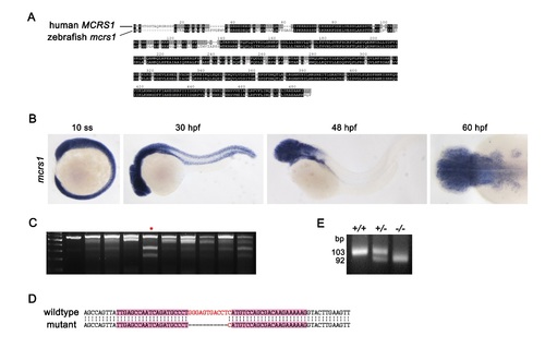
TALEN-mediated knockout of mcrs1 in zebrafish. (A) Alignment of human and zebrafish MCRS1 protein sequence (ClustaIX program). (B) Expression of mcrs1 mRNA during embryonic development was examined by whole-mount in situ hybridization (ss, somite stage; hpf, hours after fertilization). (C) T7E1 cleavage assay for the identification of zebrafish carrying a deletion in mcrs1 gene. Cleaved bands in lane 5 indicates mcrs1 het mutation. (D) Deletion of 11 base pairs in mcrs1 gene exon 4 was confirmed by DNA sequencing. (E) Genotyping of mcrs1 mutants using PCR. EXPRESSION / LABELING:
|

Phenotype of homozygous mcrs1 mutant zebrafish. (A) Homozygous mcrs1 mutants showed increased apoptotic cell death in the central nervous system. Apoptotic cells were detected by acridine orange staining at 48 and 72 hpf. Insets are magnified view of the head. (B) Injection of mcrs1 mRNA rescued excessive apoptotic cell death in mcrs1 mutant zebrafish. (C) The retinal sections from embryos at 48 and 60 hpf were stained with anti-active caspase3 antibody. (D) The retinal sections from embryos at 48 hpf were stained with anti-phospho-histone H3 antibody. (E) Whole-mount in situ hybridization of cmlc2. The establishment of left/right body axis is normal in homozygous mcrs1 mutants. PHENOTYPE:
|