
Fig. S6
- ID
- ZDB-IMAGE-160713-3
- Publication
- Lee et al., 2016 - MCRS1 associates with cytoplasmic dynein and mediates pericentrosomal material recruitment
- All Figures
- Figures for Lee et al., 2016
|
Fig. S6
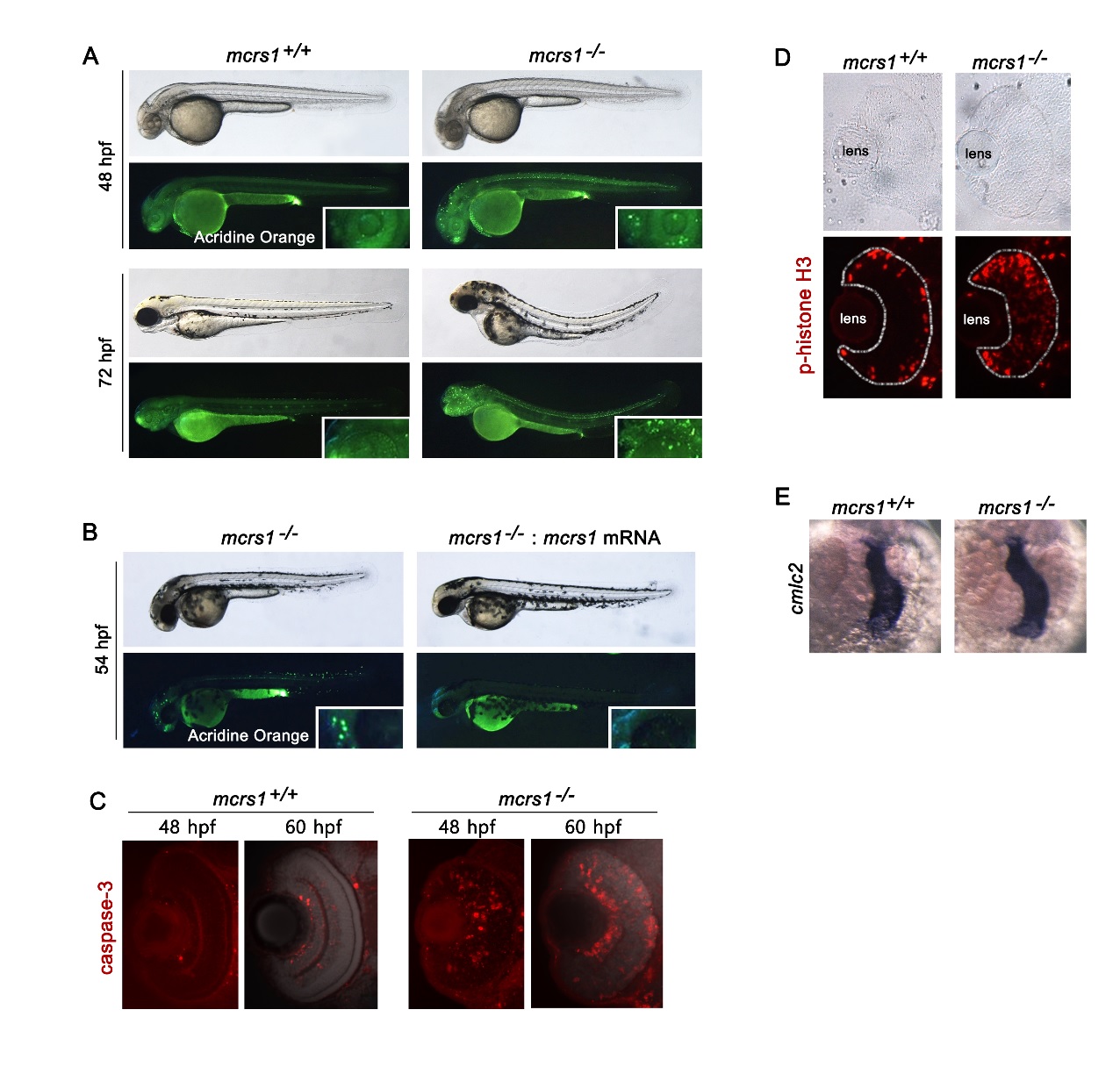
Phenotype of homozygous mcrs1 mutant zebrafish. (A) Homozygous mcrs1 mutants showed increased apoptotic cell death in the central nervous system. Apoptotic cells were detected by acridine orange staining at 48 and 72 hpf. Insets are magnified view of the head. (B) Injection of mcrs1 mRNA rescued excessive apoptotic cell death in mcrs1 mutant zebrafish. (C) The retinal sections from embryos at 48 and 60 hpf were stained with anti-active caspase3 antibody. (D) The retinal sections from embryos at 48 hpf were stained with anti-phospho-histone H3 antibody. (E) Whole-mount in situ hybridization of cmlc2. The establishment of left/right body axis is normal in homozygous mcrs1 mutants.