
- Title
-
Retinoic acid signaling controls the formation, proliferation and survival of the blastema during adult zebrafish fin regeneration
- Authors
- Blum, N., and Begemann, G.
- Source
- Full text @ Development
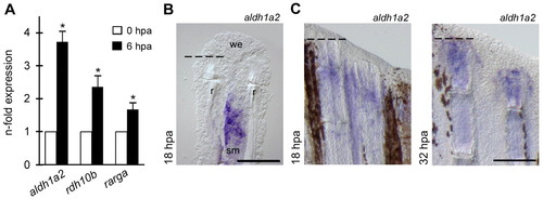
Fin amputation induces RA synthesis in the stump tissue. (A) qPCR determination of aldh1a2, rdh10b and rarga transcript levels at 6 hpa relative to uncut (0 hpa) fins. Error bars, s.e.m. *, P<0.01. (B,C) In situ hybridization on longitudinal section (B) and whole fins (C) demonstrates aldh1a2 expression in the stump mesenchyme. Note the absence of aldh1a2 transcripts in the most distal mesenchyme at 18 hpa. sm, stump mesenchyme; r, hemiray; we, wound epidermis. Dashed lines indicate amputation plane. Scale bars: 100 μm in B; 200 μm in C. EXPRESSION / LABELING:
|
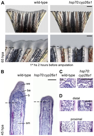
RA signaling is necessary for blastema formation. (A) Inhibition of RA signaling in hsp70:cyp26a1 fish by applying daily heat shocks (commencing 2 hours before fin amputation) results in an early and complete block to fin regeneration. (B-D) Hematoxylin-stained longitudinal sections indicate absence of blastema cells in hsp70:cyp26a1 fins at 45 hpa and lack of a distinctive basal epidermal layer. Several layers of epithelial cells seal the amputation plane, indicating normal initial wound healing. Remodeling of the stump mesenchyme adjacent to the amputation site is apparent in both wild-type and hsp70:cyp26a1 fish. (B) Overviews of stained sections. (C,D) Magnified view of the wound epidermis-mesenchyme boundary (C) and the stump mesenchyme (D). Dashed lines indicate amputation plane. hs, heat shock; b, blastema; be, basal epidermal layer; sm, stump mesenchyme; we, wound epidermis. Scale bars: 500 μm in A upper panels; 200 μm in A lower panels; 100 μm in B; 50 μm in C,D. PHENOTYPE:
|
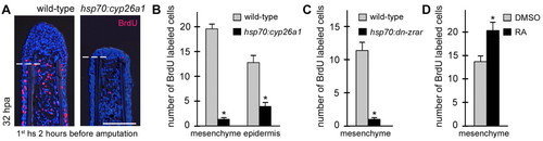
Induction of cell proliferation in the fin stump requires RA signaling. (A-C) Inhibition of RA signaling in hsp70:cyp26a1 and hsp70:dn-zrar fish results in a significant decrease in proliferating cells in the fin stump (daily heat shocks, commencing 2 hours before amputation). (A) Longitudinal sections stained for BrdU and with DAPI demonstrate a near absence of BrdU-positive cells in hsp70:cyp26a1 fins at 32 hpa. (B,C) Quantification of BrdU-labeled cells within a defined area at 32 hpa in hsp70:cyp26a1 (wild type, n=39 sections; hsp70:cyp26a1, n=44) (B) or hsp70:dn-zrar stumps (wild type, n=20; hsp70:dn-zrar, n=31) (C). (D) Exogenous RA promotes mesenchymal proliferation in the stump [RA intraperitoneal injection (IP) every 12 hours, first IP at 0 hpa]. Quantification of BrdU-labeled cells at 32 hpa in RA-treated stumps (DMSO vehicle, n=16; RA, n=18). Error bars, s.e.m. *, P<0.0001 in B,C; *, P<0.005 in D. Dashed lines indicate the amputation plane. hs, heat shock. Scale bar: 100 μm. PHENOTYPE:
|
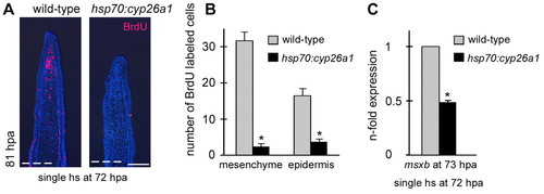
RA signaling is required for blastema proliferation. Inhibition of RA signaling in hsp70:cyp26a1 fish, when instigated during regenerative outgrowth, results in downregulation of msxb expression and loss of blastema proliferation. (A) Longitudinal sections stained for BrdU and with DAPI demonstrate absence of BrdU-positive cells in hsp70:cyp26a1 regenerates at 81 hpa after a single heat shock at 72 hpa. (B) Quantification of BrdU-labeled cells (wild type, n=17 sections; hsp70:cyp26a1, n=16). (C) qPCR determination of msxb transcript levels in hsp70:cyp26a1 regenerates relative to wild-type regenerates at 73 hpa. Error bars, s.e.m. *, P<0.0001. Dashed lines indicate amputation plane. Scale bar: 100 μm. EXPRESSION / LABELING:
|
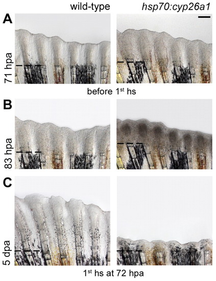
RA signaling is essential for regenerative outgrowth and maintenance of the regenerate. Inhibition of RA signaling in hsp70:cyp26a1 fish during regenerative outgrowth blocks further regeneration and abolishes maintenance of the regenerate (daily heat shocks, first heat shock at 72 hpa). (A) Before the first heat shock, regeneration in hsp70:cyp26a1 fish is indistinguishable from that of wild-type fish. (B) Ten hours after the first heat shock, the blastema of hsp70:cyp26a1 fish regenerates appears dark. (C) Two days later, regenerative outgrowth is blocked and already regenerated tissue is lost. Note the decrease in tissue distal to the amputation plane in hsp70:cyp26a1 regenerates between A and C. Dashed lines indicate amputation plane. hs, heat shock. Scale bar: 200 μm. |
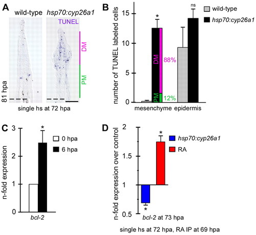
Blastema cells possess a strong RA-mediated pro-survival mechanism. (A,B) Loss of RA signaling during regenerative outgrowth causes cell death in the regenerate after a single heat shock at 72 hpa. (A) TUNEL staining on longitudinal sections at 81 hpa reveals massive cell death in the distal mesenchyme in hsp70:cyp26a1 fish. (B) Quantification of TUNEL-labeled cells in hsp70:cyp26a1 regenerates at 81 hpa (wild type, n=35 sections; hsp70:cyp26a1, n=31). Pink and green bars in B show the ratio of labeled cells between the distal and proximal mesenchyme of hsp70:cyp26a1 fish as a percentage of the total number of labeled cells in the mesenchyme. (C) qPCR determination of bcl2 transcript levels at 6 hpa relative to uncut (0 hpa) fins. (D) bcl2 transcript levels, determined by qPCR, in heat-shocked hsp70:cyp26a1 fish (control is heat-shocked wild-type fish) and RA-treated fish (control is vehicle-treated fish) relative to control fish at 73 hpa (single heat shock at 72 hpa, single RA IP at 69 hpa). Error bars, s.e.m. *, P<0.0001 in B; *, P<0.01 in C; *, P<0.001 in D; ns, not significant. Dashed lines indicate amputation plane. DM, distal mesenchyme; PM, proximal mesenchyme; hs, heat shock. Scale bar: 100 μm. |
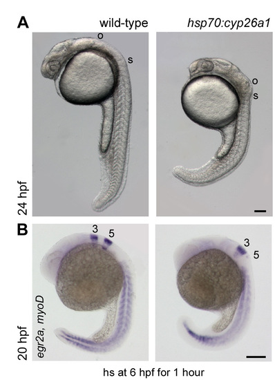
The hsp70:cyp26a1 line efficiently inhibits RA signaling. (A) Heat shock of hsp70:cyp26a1 embryos for 1 hour starting at 6 hpf results in close proximity of the first somite to the otic vesicle and a kink at the head-trunk boundary at 24 hpf, representative of complete loss of RA signaling. (B) Whole-mount in situ hybridization for egr2a, a marker of hindbrain rhombomeres 3 and 5, illustrates absence of rhombomere 5 in hsp70:cyp26a1 embryos, caused by posterior expansion of more anterior rhombomeres. hs, heat shock; o, otic vesicle; s, first somite; 3, rhombomere 3; 5, rhombomere 5. Scale bars: 100 μm. EXPRESSION / LABELING:
PHENOTYPE:
|
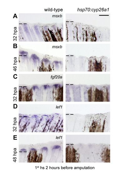
Expression of markers for the blastema and the basal epidermal layer are impaired in the absence of RA signaling. Whole-mount in situ hybridization demonstrates that neither the blastema nor the basal epidermal layer is formed or specified correctly in heat-shocked hsp70:cyp26a1 fish (daily heat shocks, first heat shock 2 hours before amputation). (A-C) In wild-type regenerates, msxb and fgf20a are strongly expressed in cells of the forming blastema at 32 or 46 hpa. Expression of both genes is undetectable in regenerates of hsp70:cyp26a1 fish. (D,E) Expression of lef1, which marks the basal epidermal layer and distal blastema cells in wild-type regenerates, is absent in hsp70:cyp26a1 fins at 32 and 48 hpa. Dashed lines indicate the amputation plane. hs, heat shock. Scale bar: 200 μm. EXPRESSION / LABELING:
|

Impaired blastema formation in RA-deficient regenerates is independent of the wound healing process. Inhibition of RA signaling in hsp70:cyp26a1 fish after wound healing has taken place results in a complete block to fin regeneration (daily heat shocks, first heat shock at 24 hpa). Dashed lines indicate the amputation plane. hs, heat shock. Scale bars: 500 μm upper panels; 200 μm lower panels. PHENOTYPE:
|
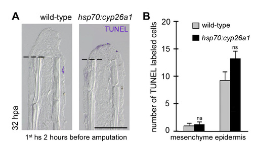
Inhibition of RA signaling does not enhance cell death during blastema formation. Inhibition of RA signaling in hsp70:cyp26a1 fish does not enhance the number of TUNEL-positive cells in the fin stump (daily heat shocks, first heat shock 2 hours before amputation). (A) Longitudinal sections stained for TUNEL show few TUNEL-positive cells in the epidermis of wild-type and hsp70:cyp26a1 fins at 32 hpa. (B) Quantification of TUNEL-labeled cells within a defined area at 32 hpa (wild type, n=32 sections; hsp70:cyp26a1, n=25). Error bars, s.e.m. ns, not significant. Dashed lines indicate the amputation plane. hs, heat shock. Scale bar: 100 μm. |
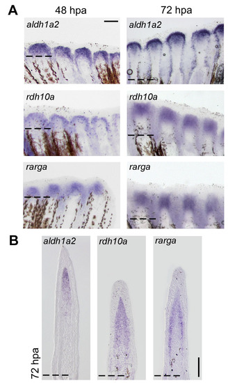
RA pathway components are highly expressed during regenerative outgrowth. Whole-mount in situ hybridization at 48 and 72 hpa (A) and in situ hybridization on longitudinal sections at 72 hpa (B) reveal expression of aldh1a2, rdh10a and rarga in the blastema. rarga and rdh10a transcripts can be detected in the whole blastema at 72 hpa, whereas aldh1a2 expression is more restricted to distal blastema cells. Dashed lines indicate amputation plane. Scale bars: 200 μm in A; 100 μm in B. EXPRESSION / LABELING:
|