
- Title
-
White adipose tissue development in zebrafish is regulated by both developmental time and fish size
- Authors
- Imrie, D., and Sadler, K.C.
- Source
- Full text @ Dev. Dyn.
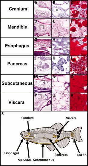
White adipose tissue (WAT) is located in distinct depots in adult zebrafish. A-R: Serial cryosections from adult (12-18 months old) zebrafish were stained with H&E (A,B,D,E,G,H,J,K,M,N,P,Q) or oil red O (C,F,I,L,O,R) for neutral lipids. The boxed region in the far left panels is magnified in the middle panels. Adipocytes have a unilocular lipid droplet (B,C asterisk) thin cytoplasm and peripheral nucleus (B,C arrow). The pancreatic acinar cells are labeled in K. Scale bar = 200 μm in the right panels and 50 μm in the middle and left panels. S: Diagram of the major sites of WAT in zebrafish adults (yellow circles). |

Orthologs of mammalian adipocyte marker genes are enriched in adult visceral, pancreatic, and esophageal adipocytes. The liver, pancreas which contains adipocytes, and visceral white adipose tissue (WAT) were dissected from three batches of three to five adult zebrafish, pooled, and processed for quantitative polymerase chain reaction (qPCR; A,B). A:amylase is a specific marker of pancreatic acinar cells and fabp10 is only expressed in hepatocytes, demonstrating the specificity of the dissected tissues. The zebrafish orthologs of acrp30, cfd, fabp11a, and pparg are expressed specifically in visceral adipose tissue and pancreas as opposed to liver. B: Bars represent the standard deviation and *indicates P < 0.05. C–L: Oil red O staining (C,H) and in situ hybridization (D-G,I-L) was carried out on consecutive serial sections of adult fish through the visceral and esophageal WAT stores. acrp30 and cfd antisense probes show positive staining in visceral (D-F) and esophageal (I-L) adipocytes coincides with positive oil red O staining in (C,H). There is some staining with both probes in esophageal muscle adjacent to the adipocytes (I,K) which is absent from sections stained with the respective senses probe (J,L). Scale bar = 50 μm. EXPRESSION / LABELING:
|

White adipose tissue (WAT) is detectable in zebrafish larvae on 12 days postfertilization (dpf). A: Representative image of a larva at 9 dpf which lacks any morphologically identifiable adipocytes. B: Rectangle is magnified to show the exocrine pancreas lacks adipocytes. C-F: On 12 dpf, the pancreas of some fish (C,E,F) but not others (D) have adipocytes (C, black asterisks) intertwined with pancreatic acinar cells. Another 12 dpf fish with serial sections stained with hematoxylin and eosin (H&E; E) or oil red O (F) show lipid-laden adipocytes. G: Time course of adipocyte appearance in over 200 zebrafish kept at the same density (10 fish/L) and fed the same amount of food. Every fish at each time point was measured for standard length (SL). Serial paraffin sections were H&E stained and scored for adipocytes. Fish where WAT was observed in any depot are represented in black dots and fish lacking any WAT are represented in white dots. Scale bar = 100 μm in A; 50 μm in B. |

Cells other than adipocytes contain lipid. A: Cryosections of adult zebrafish (12-18 months) were sectioned (30 μm) and stained in oil red O. Small lipid droplets are present in hepatocytes, skeletal muscle, and chondrocytes (black arrow heads), but not in mucous cells (black arrows), which have a similar morphology to adipocytes. Scale bar = 50 μm. B: Whole-mount oil red O staining of a 5 dpf larvae show lipid accumulation in several tissues, including the yolk, liver, jaw, and brain, but no large unilocular lipid droplets are observed in areas that coincide with regions of the fish that contain cells with adipocyte morphology. Scale bar = 1 mm. |

Adult adipocytes in the tail fin and pancreas. A-D: Cryosections of adult zebrafish (12-18 months) were sectioned (10 μm) and stained with hematoxylin and eosin (H&E; A,C) or oil red O (B,D). Sections are representative, but not consecutive. Scale bar = 50 μm. At this section thickness, oil red O staining is specific to pancreatic and tail fin adipocytes within these tissues (B,D). |

cebpα and cebpβ are expressed in adult adipocytes and liver. Quantitative polymerase chain reaction (qPCR) analysis of dissected visceral adipose tissue, exocrine pancreas and liver adult zebrafish (12-18 months). Tissue from 5-7 fish was pooled for each gender. The zebrafish cebpα and cebpβ genes are expressed in all of these tissues; cebpα in the pancreas is significantly higher compared with expression in the liver (* indicates P < 0.05). Bars represent the standard deviation. EXPRESSION / LABELING:
|