
|
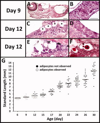
Fig. 3 White adipose tissue (WAT) is detectable in zebrafish larvae on 12 days postfertilization (dpf). A: Representative image of a larva at 9 dpf which lacks any morphologically identifiable adipocytes. B: Rectangle is magnified to show the exocrine pancreas lacks adipocytes. C-F: On 12 dpf, the pancreas of some fish (C,E,F) but not others (D) have adipocytes (C, black asterisks) intertwined with pancreatic acinar cells. Another 12 dpf fish with serial sections stained with hematoxylin and eosin (H&E; E) or oil red O (F) show lipid-laden adipocytes. G: Time course of adipocyte appearance in over 200 zebrafish kept at the same density (10 fish/L) and fed the same amount of food. Every fish at each time point was measured for standard length (SL). Serial paraffin sections were H&E stained and scored for adipocytes. Fish where WAT was observed in any depot are represented in black dots and fish lacking any WAT are represented in white dots. Scale bar = 100 μm in A; 50 μm in B.