
- Title
-
hrT is required for cardiovascular development in zebrafish
- Authors
- Szeto, D.P., Griffin, K.J.P., and Kimelman, D.
- Source
- Full text @ Development

hrT morphant phenotypes. (A) Binding positions of the morpholino antisense oligonucleotides, hrTMO(1) and hrTMO(2), to the hrT transcript. The sequence of the control-MO is also shown. (B-E) Lateral views with anterior towards the left. (B,C) The overall morphology of an uninjected (B) and hrTMO(1)-injected (C) embryos at 24 hpf. (D,E) The overall morphology of an uninjected (D) and hrTMO(2)-injected (E) embryos at 50 hpf. (F-I) Lateral view with anterior towards the top. (F,G) Heart morphology of uninjected embryo (F) and of hrTMO(1)-injected (G) embryo at 48 hpf. (H,I) Higher magnification of F,G, respectively (v, ventricle; a, atrium). Live (J,L,N,P) and green fluorescent (K,M,O,Q) pictures of embryos injected at the shield stage with: (J,K) 0.1 ng of hrT-GFP RNA; (L,M) 0.1 ng of hrT-GFP RNA plus 1.5 ng of hrTMO(1); (N,O) 0.1 ng of GFP RNA; and (P,Q) 0.1 ng of GFP RNA plus 1.5 ng of hrTMO(1). |
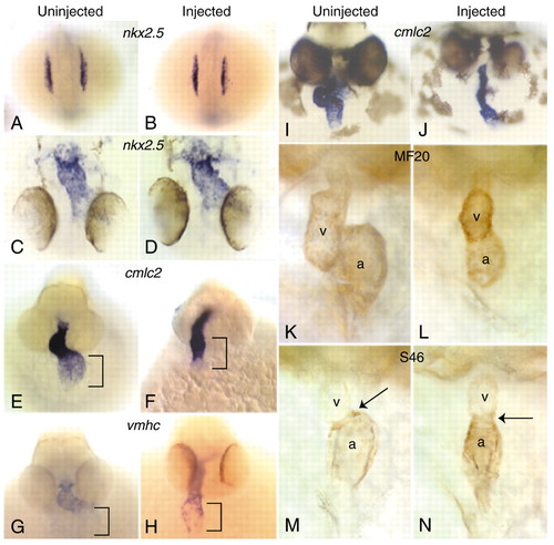
In situ and immunohistological analysis of cardiogenesis in the hrT morphants. (A-D) Dorsal views with anterior at the bottom. (A,B) Expression of nkx2.5 in uninjected (A) and hrTMO(1)-injected (B) embryos at the 12-somite stage in bilateral stripes. (C,D) Expression of nkx2.5 in the linear cardiac tube of uninjected (C) and hrTMO(1)-injected (D) embryos at 24 hpf. (E-N) Frontal views with anterior towards the top. (E,F) Cardiac expression pattern of cmlc2 in uninjected (E) and hrTMO(1)-injected (F) embryos at 33 hpf. Black bracket indicates the atrial region. (G,H) Cardiac expression patterns of vmhc in uninjected (G) and hrTMO(1)-injected (H) embryos at 33 hpf. Black bracket marks the atrial region. (I,J) Expression of cmlc2 in uninjected (I) and hrTMO(1)-injected (J) embryos at 36 hpf when cardiac looping is taking place. The injected embryo (J) has defective looping. (K,L) MF20 antibody stains the atrium and ventricle of uninjected (K) and hrTMO(1)-injected (L) embryos at 48 hpf. (M,N) S46 antibody stains the atrium of uninjected (M) and hrTMO(1)-injected (N) embryos at 48 hpf. Black arrows indicate the atrioventricular boundary. (a, atrium; v, ventricle). Injected embryos were injected with 1.5 ng of hrTMO(1). EXPRESSION / LABELING:
|

HrT regulates tbx5. (B,D,F,H) Embryos injected with 1.5 ng hrTMO(1) are compared with uninjected (A,C,E,G) embryos. (A-D) Dorsal views with anterior towards the bottom. (A,B) Expression of tbx5 at the 15-somite stage. (C,D) Expression of tbx5 at 24 hpf in the heart field. (E,F) Frontal views with anterior towards the top shows the expression of tbx5 at 33 hpf in the heart field. Note increased expression of tbx5 in hrTMO(1)-injected embryos (F). (G,H) Dorsal views with anterior to the top shows the expression of tbx5 at 33 hpf in the fin buds. (I-L) Embryos were injected with 0.2 ug of hrT-GR RNA and half were treated with dexamethasone (Dex) as indicated. (I,J) Frontal views with anterior to the top of 30 hpf embryos showing tbx5 expression in the heart. Note decreased tbx5 expression after GR-hrT induction. (K,L) Dorsal views with anterior towards the top of 30 hpf embryos, showing the expression of tbx5 in the developing fin buds. GR-hrT induction did not change the fin bud expression of tbx5. EXPRESSION / LABELING:
|
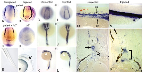
Analysis of gene expression during hematopoiesis and the formation of the trunk vasculature. (A-D) 8- to 10-somite stage embryos; posterior views with dorsal to the top. (A,B) Expression of gata1 in uninjected (A) and hrtMO(1)-injected (B) embryos. Note that injected embryos express gata1 in the most posterior region of the embryo, in contrast to uninjected embryos. (C) Expression of hrT and gata1 in uninjected embryo. (D) Expression of hrT in hrTMO(1)-injected embryo. The same expression pattern is found in uninjected embryos (not shown). Note that the hrT expression domain coincides with the region of ectopic gata1 expression in the morphants (B). (E,F) Live pictures of the posterior half of uninjected (E) and hrTMO(1)-injected (F) embryos at 36 hpf. Arrow indicates the blood pooling in the peri-anal region of the injected embryos. (G,H) Posterior views with dorsal towards the top. fli1 is expressed in a `U′-shaped pattern at 14 hpf in uninjected (G) and hrTMO(1)-injected (H) embryos. (I,J) Dorsal views with anterior towards the top. Expression of fli1 at 20 hpf. In uninjected embryos, fli1-expressing cells are at the midline (I), whereas fli1-expressing cells have not converged completely to the midline in hrT morphants (J). (K,L) Lateral views with anterior towards the top. The expression pattern of fli1 in uninjected (K) and hrTMO(1)-injected embryos at 24 hpf. White arrowhead indicates the fli1 expression in the pharyngeal primordium. (M,N) Lateral views of 24 hpf embryos with anterior towards the left. In uninjected embryos (M), fli1 expression is apparent in the dorsal aorta (da), axial vein (av) and intersegmental vessels (is), whereas hrT morphants (N) exhibit a single domain of fli1 expression in the midline of the entire trunk with no intersegmental vessels. (O,P) Transverse sections of embryos at 30 hpf stained with fli1, dorsal towards the top. In uninjected embryos (O), the formation of the dorsal aorta (black arrow) below the notochord (n) and axial vein (black arrowhead) is apparent. In the hrT morphants (P), only a single lumen is present. The black bracket indicates the developing region of the dorsal aorta and axial vein. Injected embryos were injected with 1.5 ng of hrTMO(1). EXPRESSION / LABELING:
PHENOTYPE:
|
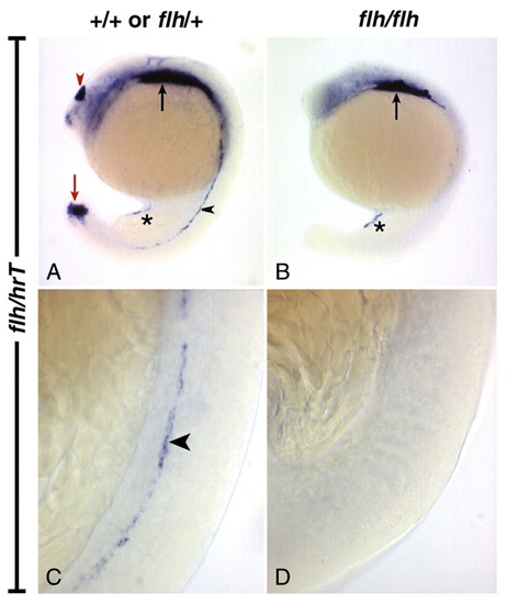
The expression of flh and hrT in flh mutant embryos. (A-D) Lateral views of 17-somite embryos with anterior towards the top, hybridized with a mixture of flh and hrT probes. In wild-type embryos or flh heterozygotes (A), flh is expressed in the tip of the tail (red arrow) and epiphysis (red arrowhead), and hrT is expressed in the dorsal aorta (black arrowhead), ventral region of the gut tube (asterisk) and in the heart (black arrow). In flh homozygotes (B), flh expression is absent and hrT expression is missing in the dorsal aorta. The gut tube and cardiac expression of hrT is not affected. (C,D) Higher-power views of the embryos in A,B, respectively. (C) The dorsal aorta expression of hrT (black arrowhead) in a wild-type or flh heterozygote. (D) The complete absence of hrT expression in the dorsal aorta of a homozygous flh embryo. EXPRESSION / LABELING:
|

Unillustrated author statements EXPRESSION / LABELING:
PHENOTYPE:
|