
- Title
-
Multiple delta genes and lateral inhibition in zebrafish primary neurogenesis
- Authors
- Haddon, C., Smithers, L., Schneider-Maunoury, S., Coche, T., Henrique, D. and Lewis, J.
- Source
- Full text @ Development
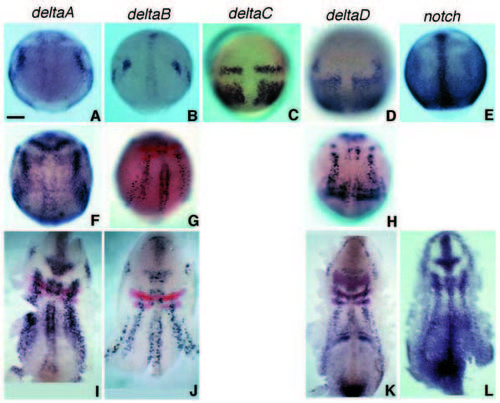
Early expression patterns of all four zebrafish delta genes and of notch seen by in situ hybridisation. Dorsal views, anterior to the top. (A-E) At 90% epiboly (9 hpf); (F-L) at bud to 1-somite stage (10-10.5 hpf). The middle row (FH) shows intact embryos, the bottom row (I-L), dissected flat mounts. Red stain in G and in I-K shows expression of krox20 and, in K, of paxb. Tissue sections (not shown) confirm that deltaA and deltaB are expressed in the epiblast, in prospective neural tissue, whereas the strong expression of deltaC and deltaD at these stages is in the prospective mesoderm. Scale bar: 100 μm. EXPRESSION / LABELING:
|

Expression of deltaA, B, C and D, notch and islet1 at the 5- somite stage (11.7 hpf), shown by in situ hybridisation with a purple blue (NBT/BCIP) detection system; embryos have also been double labelled with probes against paxb and Krox20, using a Fast-Red detection system, to provide landmarks. On the right, enlargements of the posterior hindbrain and anterior spinal-cord region are shown. Embryos are flat-mounted; anterior is to the left. tg, trigeminal ganglion; mh, midbrain/hindbrain boundary; r3, r5, rhombomeres 3 and 5; mn, motor neurons; in, interneurons; RB, Rohon-Beard (sensory) neurons; som, prospective somite. Scale bars: 200 μm (whole embryos), 100 μm (details). EXPRESSION / LABELING:
|
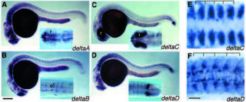
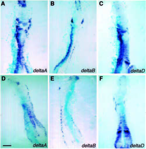
(A-D) Expression of deltaA, deltaB, deltaC and deltaD in whole mounts at 24 hpf. Note expression of deltaC and deltaD in the presomitic mesoderm (psm) of the tail bud and in recently formed somites. deltaC is now also strongly expressed in the retina (r), while deltaA, deltaB, and deltaD are expressed in scattered subsets of cells in the brain and spinal cord (sc). (E,F) Expression of deltaC and deltaD in recently formed somites at the 10-somite stage (14 hpf). Trunk region of flat-mounted embryo, anterior to the left. Note that deltaC is expressed in the anterior part of each somite, deltaD in the posterior part. Scale bars: 200 μm (A-D), 50 μm (E,F). EXPRESSION / LABELING:
|
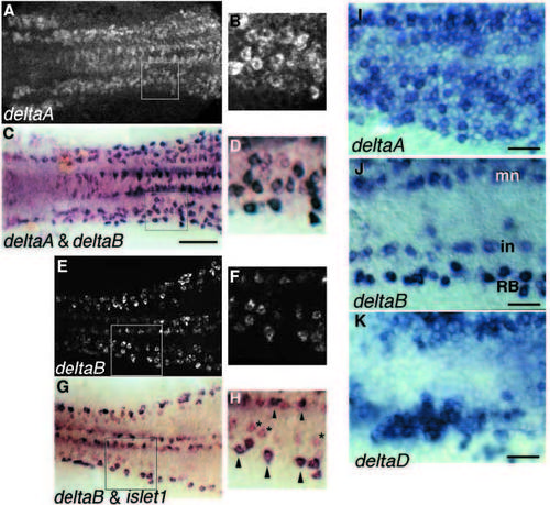
(A-D) Coexpression of deltaA and deltaB in the same cells in the neural plate at the 5-somite stage. The boxed regions on the left are shown enlarged on the right (B,D). The cells expressing deltaB are generally the same that express deltaA most strongly. The embryo was first stained for deltaA expression, revealed by in situ hybridisation using fluorescent Fast Red detection and was viewed intact by epifluorescence, using the confocal microscope to construct an extended-focus image. The same specimen was then hybridised with a probe for deltaB, revealed in purple with NBT/BCIP, and was flatmounted and photographed with bright-field optics. Sequential imaging was used because the dark NBT/BCIP stain often obscures the Fast Red fluorescence. (E-H) Coexpression of deltaB and islet1 in the neural plate at 5 somites, shown by double in situ hybridisation. deltaB in red (fluorescence in upper panel (E,F), bright field in lower panel (G,H)), islet1 in blue-black (bright field, lower panel). Primary motor neurons and Rohon-Beard cells express both genes (arrowheads); cells expressing deltaB but not islet1 are probably primary interneurons (asterisks). (I-K) Details of the prospective anterior spinal region at the 5-somite stage, showing the diffuse but uneven expression of deltaA and deltaD in large patches of contiguous cells, and the more restricted expression of deltaB in scattered, isolated cells. In each case, the left side of the neural plate is shown, with midline at the top and anterior to the left of the picture. Scale bars: 100 μm (A-H), 50 μm (I-K). EXPRESSION / LABELING:
|
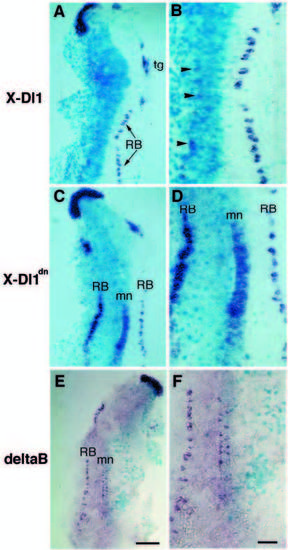
(A-D) Effects on primary neurogenesis following injection of X-Delta1 or X-Delta1dn RNA, together with lacZ RNA as a marker, into one blastomere. Flat-mounted embryos at 5- to 6-somite stage, with islet1 expression in purple-blue, lacZ marker in sky-blue; lowmagnification views on the left, details on the right; note that the Xgal treatment of the embryos for detection of lacZ product results in fainter in situ hybridisation stainings than in Figs 2-5. X-Delta1 inhibits production of all classes of islet1-positive primary neurons in the injected (sky-blue) region; X-Delta1dn does the opposite. Arrowheads in B indicate a few Rohon-Beard cells that have been formed despite the injected X-Delta1. Note that convergence movements and folding of the neural plate lead to cell mixing in the midline, so that motor neuron production appears affected uniformly on both sides of the midline. tg, trigeminal ganglion; mn, motor neurons; RB, Rohon-Beard neurons. (E,F) Effects on primary neurogenesis following injection of deltaB RNA, together with lacZ RNA as a marker, into one blastomere. Flat-mounted embryos at 5- to 6-somite stage, with islet1 expression in purple-blue, lacZ marker in sky-blue; low magnification views on the left, details on the right. Where deltaB RNA is present, production of islet1-positive primary neurons is inhibited. Scale bars: 200 μm (A,C,E), 100 μm (B,D,F). |

Effects on Mauthner cell production following injection of X-Delta1 RNA or X-Delta1dn RNA into one blastomere. Embryos were fixed at 30 hpf and stained with 3A10 antibody. (A) Normal control; (B) X-Delta1 injection, leading to loss of the Mauthner cell on one side of the brain; (C-D) X-Delta1dn injections, giving supernumerary Mauthner cells (arrowheads). Scale bars: 100 μm (A-C), 50 μm (D). EXPRESSION / LABELING:
|
Effects on expression of delta genes in the neural plate following injection of X-Delta1 or X-Delta1dn RNA, together with lacZ RNA as a marker, into one blastomere. Flat mounts at 5- to 6- somite stage, with expression of deltaA, deltaB and deltaD in purpleblue, lacZ marker in sky-blue. Where X-Delta1 RNA is present, expression of all three delta genes is inhibited; where X-Delta1dn RNA is present, they are all overexpressed. Note in A-C that dramatic effects of X-Delta1dn are seen even in regions where there is normally only low-level expression of endogenous delta genes. Scale bar, 200 μm. |