
- Title
-
Expression of zebrafish nk2.2 is influenced by sonic hedgehog/vertebrate hedgehog-1 and demarcates a zone of neuronal differentiation in the embryonic forebrain
- Authors
- Barth, K.A. and Wilson, S.W.
- Source
- Full text @ Development
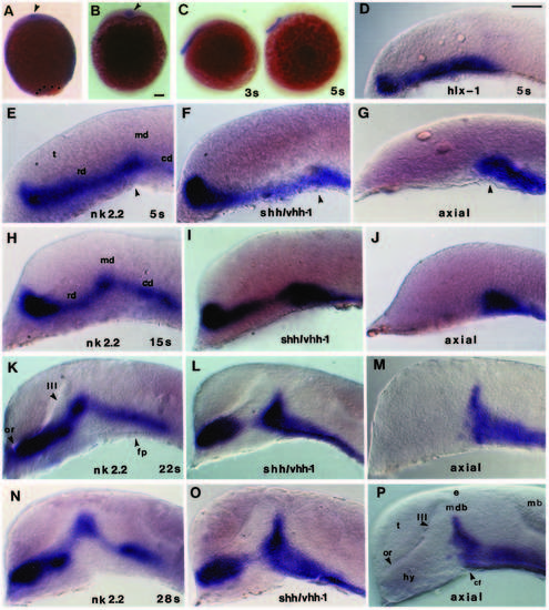
Comparison of the developmental time course of nk2.2 expression in the rostral brain with that of shh/vhh-1 and axial. Whole-mount embryos hybridised with antisense RNA to nk2.2, shh/vhh-1, axial or hlx-1. Lateral views (except A,B) are shown with rostral to the left. In DY, the skin, yolk and eyes have been removed. (A,B) Frontal views (with dorsal up) showing nk2.2 expression (arrowheads) at 95% epiboly (9.5h) (A) and bud/1s (10h) stage (B). Dots outline the yolk plug in A. (C) Lateral view of nk2.2 expression at 3 somites (11h) and 5 somites (11.7h). (D) hlx-1 expression in the forebrain of a 5 somites (11.7h) embryo. (E-Y) Comparison of rostral brain expression domains of nk2.2 (E,H,K,N,Q,T,W), shh/vhh-1 (F,I,L,O,R,U,X) and axial (G,J,M,P,S,V,Y) from 5 somites (11.5h) to 44-48h. The arrowheads in E-G indicate a small groove in the mid-diencephalon at which the cephalic flexure will later form. Arrowhead in Q indicates the gap between rostral and caudal nk2.2 expression domains. In V, the embryo was also labelled with an antisense RNA probe to wnt1 which is expressed in cells beneath the epiphysis (Macdonald et al., 1994). Abbreviations: cb, cerebellum; cf, cephalic flexure; e, epiphysis; fp, floorplate; hy, hypothalamus; mb, midbrain; mdb, mid-diencephalic boundary; or, optic recess; p; anlage of the anterior pituitary; rd and cd, rostral and caudal domains of nk2.2 expression; t, telencephalon; te, tegmentum; III, third ventricle. Scale bar=100 μm EXPRESSION / LABELING:
|
|
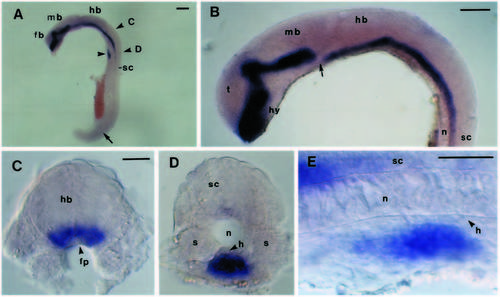
nk2.2 expression in the hindbrain and in a group of cells ventral to the notochord. Lateral views (A,B,E) with rostral to the left and transverse sections (C,D) of 20-22 somites (19-20h) embryos from which the eyes and yolk have been removed. The alkaline phosphatase colour reaction was developed 5-6 times longer than usual to reveal weak expression. The approximate levels of the sections shown in C and D are indicated in A. (A) Low magnification view of the entire embryo. The arrow indicates very faint staining in the caudal spinal cord, and the arrowhead points to the group of cells shown in D and E. (B) nk2.2 expression in the brain. The arrow indicates the discontinuity in the band of nk2.2-expressing cells. (C) Transverse section through the caudal hindbrain revealing expression in cells adjacent to the floorplate. (D) Transverse section near the hindbrain/spinal cord junction showing nk2.2 expression in cells beneath the notochord and hypochord (arrow). (E) Lateral view of the same group of cells as (D). Abbreviations: fb, forebrain; fp, floorplate; h, hypochord; hb, hindbrain; hy, hypothalamus; mb, midbrain; n, notochord; s, somite; sc, spinal cord; t, telencephalon. Scale bar: A,B=100 μm, C-E=20 μm. 
EXPRESSION / LABELING:
|
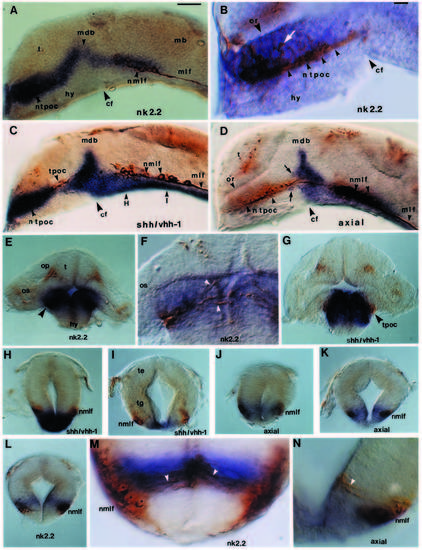
Gene expression boundaries of nk2.2, shh/vhh-1 and axial demarcate sites of neuronal differentiation and axogenesis in the forebrain and midbrain. Embryos are hybridised with nk2.2 (A,B,E,F,L,M), shh/vhh-1 (C,G-I), axial (D,J,K,N) antisense RNA and HNK-1 antibody (brown labelling of neurons and axons). (A-D) Lateral views of sagittal hemisections with rostral to the left and eyes removed. (A) nk2.2 expression with respect to the nTPOC and nMLF. The dark blue alkaline phosphatase reaction product masks the nTPOC in A and C. (B) High magnification of nk2.2 expression with respect to the nTPOC. The arrowheads indicate the course of the axons in the TPOC, and the white arrow indicates HNK1 labelling within the nk2.2 expression domain. (C,D) Correlation of shh/vhh-1 (C) and axial (D) expression domains with the locations of the nTPOC and nMLF. The arrows in D indicate a few axial-expressing cells dorsal and ventral to axons in the TPOC and the positions of the sections shown in H and I are indicated in C. (E-N) Transverse sections. (E-G) nk2.2 (E,F) and shh/vhh-1 (G) expression at the level of the nTPOC. The arrowheads in F indicate immunoreactive processes within the nk2.2 expression domain. (H-J) shh/vhh-1 (H,I), axial (J,K,N) and nk2.2 (L,M) expression at the level of the nMLF (H and J are through the rostral part of the nucleus and I,K and L are through the caudal part of the nucleus). The arrowheads in M and N indicate immunoreactive processes connecting to the ventricle. The section shown in M is from a slightly older embryo than in L. Abbreviations: cf, cephalic flexure; hy, hypothalamus; mb, midbrain; mdb, mid-diencephalic boundary; nMLF and MLF, the nucleus of the medial longitudinal fasciculus and its associated tract; nTPOC and TPOC, the nucleus of the tract of the postoptic commissure and its associated tract; or, optic recess; t, telencephalon. Scale bars for A,C,D,E, G-L and B,F,M,N=100 μm. |
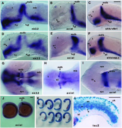
Injection of shh/vhh-1 RNA results in elevated and ectopic expression of nk2.2, axial and shh/vhh-1. Whole-mount embryos with rostral to the left. (A-F) Lateral views of 24h shh/vhh-1-injected embryos showing nk2.2 (A,D), axial (B,E) and shh/vhh-1 (C,F) expression. Eyes have been removed. (D-F) Examples of embryos affected more severely than those in A-C. (G,H) Dorsal views showing the gap in nk2.2 expression at the MDB of uninjected wild-type embryos (G) and the complementary expression of axial at the same location (H). The eyes are removed in H. (I) Dorsal view of expanded axial expression domain at the MDB in an shh/vhh-1-injected embryo. (J) Lateral view of axial expression in an control (left) and shh/vhh-1-injected 12 somite embryos. (K) Detection of b-galactosidase activity (blue) in embryos injected with b- galactosidase-encoding RNA. Some embryos were also examined for nk2.2 expression (eg. dark blue label in the forebrain of embryo at bottom left). (L) Higher magnification of the tail region of embryo seen bottom right in K. Blue cells are positive for b-galactosidase. Abbreviations: bl, blood; cf, cephalic flexure; h, hypochord; hy, hypothalamus; mb, midbrain; mdb, mid-diencephalic boundary; n, notochord; or, optic recess; sc, spinal cord; sk, skin; t, telencephalon; y, yolk; III, third ventricle. Scale bar=100 μm. |
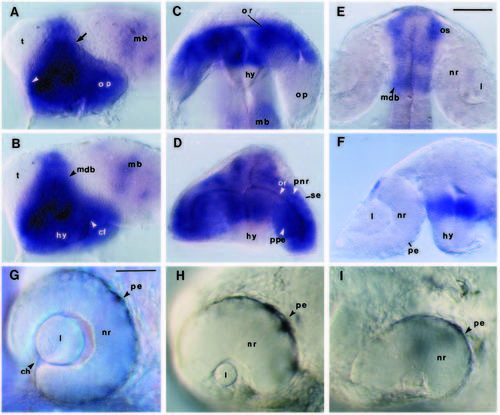
Ectopic expression of nk2.2 in the optic primordia of shh/vhh-1-injected embryos correlates with impaired eye development. (A-D) Whole-mount 22-24h shh/vhh-1-injected embryos hybridised with antisense RNA to nk2.2. (A,B) Lateral views showing ectopic nk2.2 expression throughout the optic primordia. (A) Focussed at the level of the eye and (B) focussed through the eye and onto the brain. The white arrowhead indicates the normal position of the optic stalk and the arrow indicates the dorsocaudal limit of fusion of the optic primordia to the brain. (C) Ventral view of an shh/vhh-1-injected embryo with ectopic nk2.2 expression in the anterior part of the optic primordia. (D) Frontal view of an shh/vhh-1-injected embryo with nk2.2 expression throughout the optic primordia. (E,F) Dorsal (E) and frontal (F) views of nk2.2 expression in normal 22-26 somites (20-22h) embryos. (G-H) Eye morphology in living normal (G) and shh/vhh-1-injected (H,I) 30h embryos. The lens is reduced in H and absent in I. Ventrorostral eye development and pigment formation is affected in both embryos. Abbreviations: cf, cephalic flexure; ch, choroid fissure; hy, hypothalamus; l, lens; mb, midbrain; mdb, mid-diencephalic boundary; nr, neural retina; op, optic primodia; or, optic recess; os, optic stalk; pe, pigment epithelium; pnr, presumptive neural retina; ppe, presumptive pigment epithelial layer; se, surface ectoderm; t, telencephalon. Scale bar: A-F=100 μm, G-I=50 μm. |
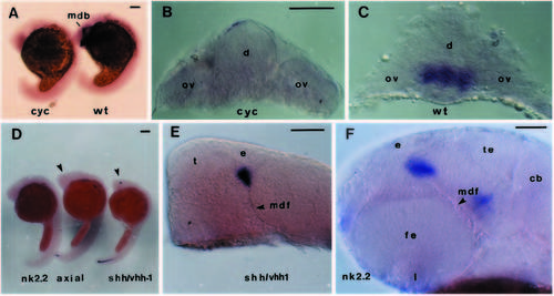
Expression of nk2.2, shh/vhh-1 and axial in homozygous mutant cyclops embryos. Lateral views (A,D,E,F) with rostral to the left, and transverse sections (B,C) of embryos hybridised with nk2.2, axial or shh/vhh-1 anti-sense RNA. (A-C) nk2.2 expression in 18 somite (18h) wild-type and cyclops mutant embryos. The transverse sections shown in B and C are at the level of the diencephalon. The small dark patch on the dorsal surface of the embryo in B is an artefact. (D) 30 somites (24h) cyclops mutant embryos hybridised with antisense RNA to nk2.2, axial and shh/vhh-1. The arrowheads indicate a small cluster of axial and shh/vhh-1-expressing cells in the forebrain. (E) shh/vhh-1 expression at the dorsal tip of the mid-diencephalic furrow of a 30 somite (24h) cyclops mutant embryo. (F) nk2.2 expression at the tip of the middiencephalic furrow of a 40-44h cyclops mutant embryo. Abbreviations: cb, cerebellum; e, epiphysis; fe, fused eye; l, lens; mdb, middiencephalic boundary; mdf, mid-diencephalic furrow; ov, optic vesicle; t, telencephalon; te, tectum. Scale bar=100 μm. EXPRESSION / LABELING:
|