
Fig. 4
- ID
- ZDB-FIG-140226-40
- Publication
- Barth et al., 1995 - Expression of zebrafish nk2.2 is influenced by sonic hedgehog/vertebrate hedgehog-1 and demarcates a zone of neuronal differentiation in the embryonic forebrain
- Other Figures
- All Figure Page
- Back to All Figure Page
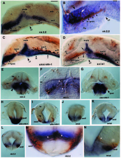
Gene expression boundaries of nk2.2, shh/vhh-1 and axial demarcate sites of neuronal differentiation and axogenesis in the forebrain and midbrain. Embryos are hybridised with nk2.2 (A,B,E,F,L,M), shh/vhh-1 (C,G-I), axial (D,J,K,N) antisense RNA and HNK-1 antibody (brown labelling of neurons and axons). (A-D) Lateral views of sagittal hemisections with rostral to the left and eyes removed. (A) nk2.2 expression with respect to the nTPOC and nMLF. The dark blue alkaline phosphatase reaction product masks the nTPOC in A and C. (B) High magnification of nk2.2 expression with respect to the nTPOC. The arrowheads indicate the course of the axons in the TPOC, and the white arrow indicates HNK1 labelling within the nk2.2 expression domain. (C,D) Correlation of shh/vhh-1 (C) and axial (D) expression domains with the locations of the nTPOC and nMLF. The arrows in D indicate a few axial-expressing cells dorsal and ventral to axons in the TPOC and the positions of the sections shown in H and I are indicated in C. (E-N) Transverse sections. (E-G) nk2.2 (E,F) and shh/vhh-1 (G) expression at the level of the nTPOC. The arrowheads in F indicate immunoreactive processes within the nk2.2 expression domain. (H-J) shh/vhh-1 (H,I), axial (J,K,N) and nk2.2 (L,M) expression at the level of the nMLF (H and J are through the rostral part of the nucleus and I,K and L are through the caudal part of the nucleus). The arrowheads in M and N indicate immunoreactive processes connecting to the ventricle. The section shown in M is from a slightly older embryo than in L. Abbreviations: cf, cephalic flexure; hy, hypothalamus; mb, midbrain; mdb, mid-diencephalic boundary; nMLF and MLF, the nucleus of the medial longitudinal fasciculus and its associated tract; nTPOC and TPOC, the nucleus of the tract of the postoptic commissure and its associated tract; or, optic recess; t, telencephalon. Scale bars for A,C,D,E, G-L and B,F,M,N=100 μm. |