Image
Figure Caption

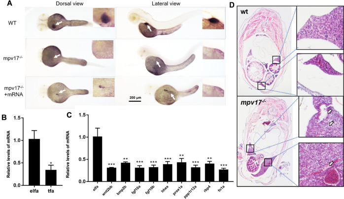
Fig. 5 The development of the liver was abnormal in the <italic>mpv17</italic><sup><italic>−/−</italic></sup> zebrafish.

A In situ hybridization with RNA probe tfa at 3 dpf. Wild-type was in the upper, mpv17−/− in the middle, and the lower was the mpv17−/− larvae rescued with mpv17 mRNA. B In situ hybridization of larvae at 5 dpf using tfa probe. C The expression level of genes tfa. Error bars were indicated with ±SEM, *p< 0.05. D The expression levels of genes involved in liver development. Error bars were indicated with ±SEM, **p< 0.01, ***p< 0.005. E H&E staining paraffin-embedded sections of the liver in 1-month-old zebrafish. The black frames were enlarged as shown on the right.

Figure Data
Acknowledgments
This image is the copyrighted work of the attributed author or publisher, and ZFIN has permission only to display this image to its users. Additional permissions should be obtained from the applicable author or publisher of the image. Full text @ Cell Death Discov