
Fig. S8
|
Fig. S8
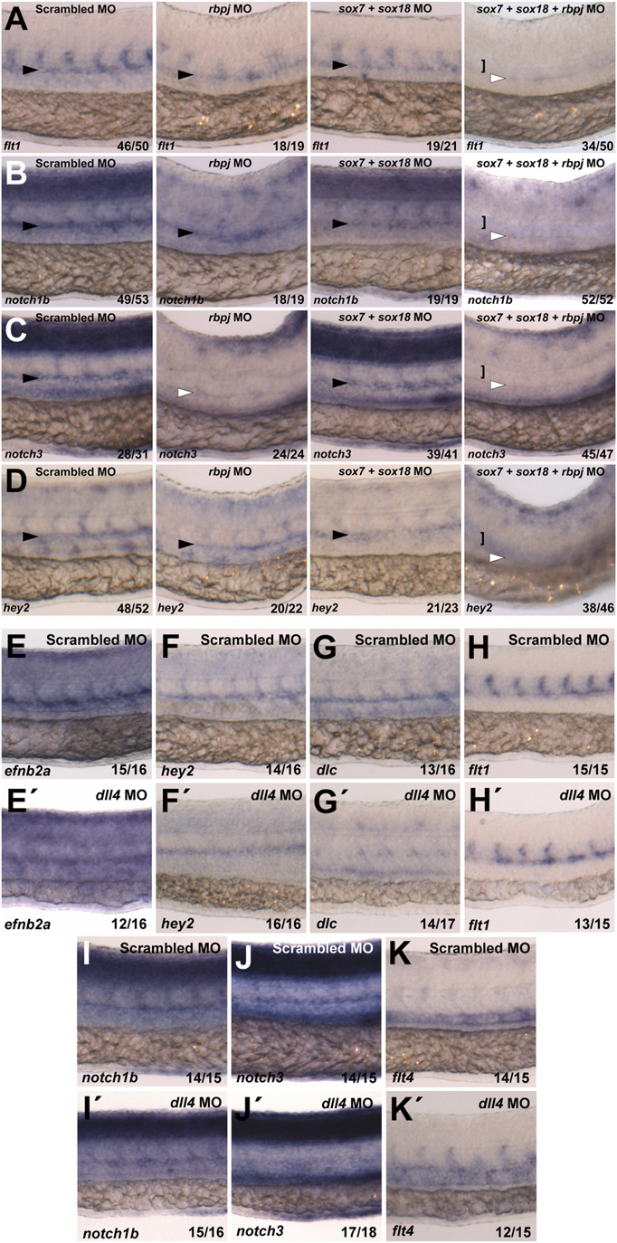
(A–D) Whole-mount in situ hybridization of arterial markers on control, rbpj, double sox7;sox18, and triple sox7;sox18;rbpj MO at 26 hpf. Triple sox7; sox18;rbpj knockdown results in the loss of all of the arterial markers analyzed. Black arrowhead shows dorsal aorta. White arrowhead indicates where dorsal aorta should be. Black arrow indicates posterior cardinal vein, and white bracket indicates single axial vessel/expanded posterior cardinal vein. Black bracket indicates notochord. Probes used are depicted in the bottom left of each picture. Values on the bottom right indicate number of embryos with the predominant and displayed phenotype/total number of embryos analyzed. In all cases, 0.125 pmol of sox7 and sox18 and 0.15 pmol of rbpj MO were used. (E–K) Whole-mount in situ hybridization of arterial (A–F) and venous (G) markers on control and dll4 MO-injected WT zebrafish embryos at 26 hpf. Probes are depicted in the bottom left. Values on the bottom right indicate number of embryos with the predominant and displayed phenotype per total number of embryos. Six nanograms of dll4 MO were used.