
Fig. 3
|
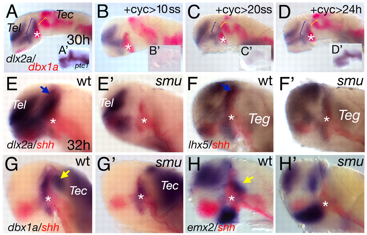
Fig. 3 The MDT is reduced in embryos deficient for Hh signalling. (A) Inhibition of Hh signalling by cyclopamine treatment (70 µm) from 10-somites to 30 hours leads to a strong reduction of dlx2a expression anterior to the ZLI and dbx1a posterior to the ZLI (B). ptc1 expression is not detectable in these embryos (B'). Weaker phenotype observed with treatment between 20 somites and 30 hours (C; dlx2a-positive prethalamus is marked by blue bracket and dbx1a by yellow bracket), although ptc1 expression is still undetectable (C'). After 24 hpf, inhibition of Hh signalling has no detected effect (D) compared with wild-type siblings (A). Asterisks indicate the ZLI (A-D). Analysis of the smu phenotype reveals Hh dependency for gene expression in the MDT (E-H'). dlx2a is not detectable in the prethalamus (E,E', blue arrow), lhx5 expression is downregulated (F,F', blue arrow), dbx1a is absent form the thalamus (G,G'; yellow arrow), in contrast to the ZLI (asterisk). Similarly, emx2 is downregulated (H,H'; yellow arrow). Tec, tectum; Teg, tegmentum; Tel, telencephalon.