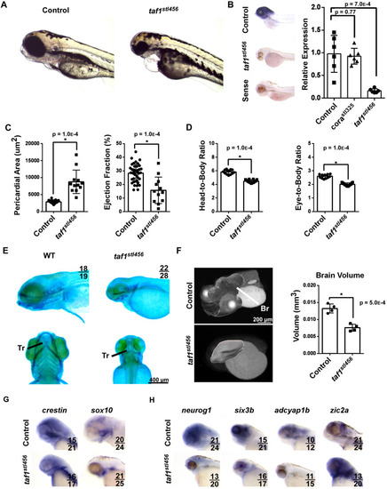
taf1 deletion recapitulates the corazoncito phenotype. (A) Brightfield images of control and taf1stl456/stl456 embryos at 96 hpf. (B) taf1 mRNA expression measured by in situ hybridization (left) and RT-PCR (right). N=6 per experimental group. (C) Quantification of pericardial area (left) and ejection fraction (right) in control (n=30) and taf1stl456/stl456 (n=12) embryos at 96 hpf. (D) Quantification of head-to-body ratio (left) and head-to-eye ratio (right) in control (n=21) and taf1stl456/stl456 (n=20) embryos at 96 hpf. (E) Alcian Blue staining of control and taf1 knockout (KO) embryos. (F) X-ray microscopy-generated images of control and taf1 KO embryos at 96 hpf (left). Quantification of brain volume (right). N=4 per experimental group. Br, brain (red), heart (blue). (G) Whole-mount in situ hybridization of crestin and sox10 mRNA expression in control and taf1 KO embryos at 96 hpf. (H) Whole-mount in situ hybridization of neuronal (neurog1) and brain region markers (six3b, Adcyap1b, zic2a).
|
