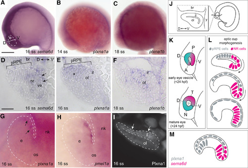
sema6d and plxna1a are expressed in complementary domains in the early eye vesicle. A, Lateral view of a 16-ss zebrafish embryo shows sema6d transcript in the eye vesicle, and regions of the head and trunk. B, C, Embryos processed for RNA ISH reveal transcript present in the optic vesicle at the 14-ss (plxna1a) and 18-ss (plxna1b) stage. D–F, Transverse sections (line in A) through the brain and eye. sema6d transcript is expressed in the ventral (future temporal) domain of the inner eye vesicle leaflet (arrows) and neural retina (outer leaflet), but is absent from the dorsal (future nasal), presumptive RPE progenitor (pRPE) domain (bar; D). plxna1a mRNA is present in the pRPE domain (bar) and faintly in the dorsal neural retina (asterisk), but absent from the ventral inner and outer eye vesicle leaflet (E). plxna1b is expressed in scattered cells of the outer leaflet of the developing optic cup (F). G, H, Eye vesicle (viewed dorsally) at the 16 ss processed for whole-mount ISH with antisense riboprobes for plxna1a (arrows in G) or the RPE marker, pmel1a (H). I, Plxna1-like immunoreactivity is present at the 16 ss in the pRPE domain (arrows). J–L, Schematics of the eye vesicle (J), the early embryo axes with respect to the eye (K), and optic cup morphogenesis (L); note that as the eye rotates alongside brain development, early ventral retina tissue (pink) becomes mature temporal tissue. M, Schematic of plxna1a and sema6d mRNA expression in the 16-ss eye vesicle. Scale bars: 300 μm (A) and 50 μm (D). br: brain, D: dorsal, e: eye, il: inner leaflet, N: nasal, nk: neural keel, nr: neural retina, ol: outer leaflet, os: optic stalk, pRPE: presumptive RPE, RPE: retinal pigment epithelium, T: temporal, V: ventral, ve: ventricle.
|
