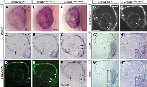
Disrupted RPE morphogenesis in sema6d mutants. A–C, Morphogenesis of the RPE is disrupted in sema6dca302 and sema6dca303 eyes as compared with wild-type, with mutants displaying ectopic bhlhe40 staining apparent through the transparent lens (white arrowhead in B, C). In the bottom right of panels are the number of embryos of the total analyzed that exhibited either a WT (A) or a disrupted (B, C) expression pattern. A’–C’, F, Transverse sections reveal full expansion of the bhlhe40+ RPE to abut the nasal lens (black arrows), while the RPE of the temporal eye fails to reach the lens in mutants (compare red arrows). RPE cells are expanded in the inner leaflet of wild-type embryos, but cuboidal in mutants (compare red asterisks). In most mutants the bhlhe40+ RPE not only failed to abut the lens, but bhlhe40 was ectopically expressed in the lateral portion of the temporal neural retina (white arrowheads in B’, C’), while in some sema6d mutants, bhlhe40 expression was restricted entirely to the inner eye vesicle leaflet (red arrow in F). D, E, Wild-type (D) and sema6dca302 mutant (E) embryos bred on a Tg(tfec:EGFP) background to label RPE progenitors. Mutants display ectopic EGFP expression in the temporal neural retina (arrowheads in E) and EGFP+ RPE cells in the temporal (early ventral) inner leaflet are cuboidal and not extended as in wild-type (compare arrows in D, E). G, H, Transverse sections of 22-hpf eyes of wild-type (G–G’’) and sema6dca302 (H–H’’) mutant embryos on a Tg(tfec:EGFP) background that were processed for GFP immunohistochemistry (G, H) and RNA ISH for the temporal neural retina marker foxd1 (G’, H’). Blended images (G’’, H’’). EGFP+ cells that co-express foxd1 are present in the distal temporal neural retina of both wild-type and mutant eyes (arrowheads in G, G’’, H, H’’). A few foxd1+ cells in the wild-type inner leaflet (asterisk in G’) have not yet moved around the distal rim, with many more present in the mutant inner leaflet (asterisk in H’). Scale bars: 50 μm (A, A’). D: dorsal, il: inner leaflet, L: lens, N: nasal, ol: outer leaflet, T: temporal, V: ventral.
|
