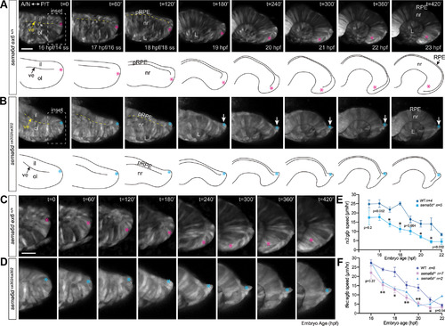
Ventral inner leaflet cells fail to move appropriately around the distal rim during optic cup morphogenesis. A, B, Confocal optical sections, and their corresponding schematics, acquired from the nasal/temporal plane between the 14 ss and 23 hpf of wild-type and sema6d mutant Tg(rx3:GFP) eyes (labels all eye progenitors). Dotted yellow line(s) indicates the separation between the inner and outer leaflets. C, D, Higher magnification of inset boxes (early ventral/future temporal retina) in A, B, respectively. In wild type (A), ventral progenitors move around the distal rim of the optic cup and come to reside in the temporal neural retina at 23 hpf. In contrast, sema6d mutants display temporal defects (follow arrow in B). While a ventral inner leaflet cells in wild type (C) moves around the distal rim (pink asterisk), a corresponding cell in the sema6d mutant (D) begins to move toward the distal rim, but stalls and, together with other progenitors, protrudes from the inner leaflet (blue asterisk). E, F, Mean of the speed of individual cells in each wild-type and sema6d mutant embryo tracked over the period of optic cup morphogenesis. Tracked were ventral neural progenitors located at the distal eye vesicle tip in the Tg(rx3:GFP) background (E) and brightly EGFP labeled presumptive RPE cells in the Tg(tfec:EGFP) background (F). In both reporter lines, sema6d mutant cells move more slowly than their wild-type counterparts, and stall at ∼18–20 hpf. ns are number of embryos assayed (2–7 cells/embryo), and error bars are SEM. Mean speeds at individual time points were compared statistically between wild-type and heterozygous embryos by a Mann–Whitney U test (p < 0.05; E, F). A: anterior, il: inner leaflet, L: lens, N: nasal, nr: neural retina, ol: outer leaflet, P: posterior, pRPE: presumptive RPE, RPE: retinal pigment epithelium, T: temporal, ve: ventricle.
|
