|
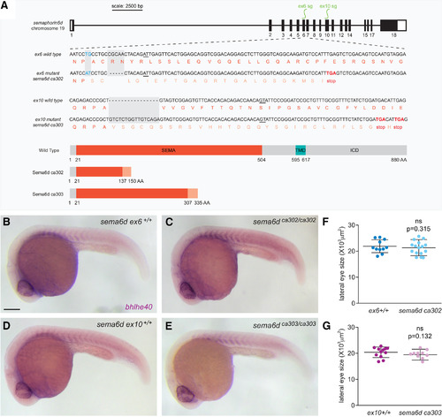
sema6d mutants do not display gross morphologic defects. A, Schematic of the two sema6d mutant alleles; sema6dca302 mutants contain a five-base pair deletion and two-base pair mismatch in exon6, and sema6dca303 mutants contain a 16-base pair insertion in exon10, both predicted to generate proteins that truncate prematurely within the SEMA domain and result in the loss of the transmembrane domain (TMD) and the intracellular domain (ICD). B–E, Lateral views of whole-mount embryos processed for bhlhe40 ISH, revealing normal gross embryonic morphology and somite labeling in both mutant alleles with respect to their wild-type siblings at 24 hpf. F, G, Eye area measured from lateral images of sema6d mutants and their respective wild-type siblings at 24 hpf [unpaired t test, df = 26 (F), df = 21 (G); error bars are SD]. Scale bar: 200 μm.
|