|
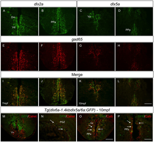
Co-expression of <italic>dlx</italic> paralogs with markers of GABAergic neurons in adult zebrafish.Double fluorescence ISH of transverse sections of the forebrain showing expression of dlx2a (A-B) and dlx5a (C-D) in green and expression of gad65 in red (E-H). Anatomical parts indicated. Merged images showing co-localization of dlx and gad65 in yellow [I-L] (N = 4 for dlx2a and dlx5a). Double IHC with Tg(dlx6a-1.4kbdlx5a/6a:GFP) and Calretinin or Calbindin shows co-localization, indicated by white arrows [N-P] (N = 6 for Calbindin and Calretinin). Merged images were created with ImageJ(32) software. Calret.: Calretinin and Calb.: Calbindin. Dm.: medial zone of dorsal telencephalic area; PGZ.: periventricular gray zone; PPa.: anterior part of parvocellular preoptic nucleus; PPp.: posterior part of parvocellular preoptic nucleus; V.: ventral telencephalic area; Vd.: dorsal nucleus of V.; Vp.: parvocellular nucleus of V.;Vs.: supracommissural nucleus of V.; Vv.: ventral nucleus of V. Scale bar: 400μm.
|