
Fig. 1
- ID
- ZDB-FIG-160923-1
- Publication
- Qiu et al., 2016 - Embryonic hematopoiesis in vertebrate somites gives rise to definitive hematopoietic stem cells
- Other Figures
- All Figure Page
- Back to All Figure Page
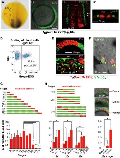
Stage- and position-dependent hematogenic activity of somites. (A) Double in situ hybridization patterns of scl (red) and eos (black/blue) in a dorsally viewed Tg(foxc1b:EOS) embryo at the 10s stage. (B) EOS protein fluorescence in somites and paraxial mesoderm in a laterally viewed Tg(foxc1b:EOS) embryo at the 10s stage. (C and C′) Double fluorescence in situ hybridization patterns of eos (red) and gata1a (green) in a Tg(foxc1b:EOS) embryo at the 10s stage. The confocal image of trunk region was dorsally viewed (C) with an optical cross section showed in C′. (D) A representative FACS result of green-EOS+ blood cells from 10 Tg(foxc1b:EOS) embryos. The average from three independent experiments was shown in parenthesis. (E and F) green-EOS in five pairs of somites in Tg(foxc1b:EOS;fli1a:gfp) embryos was converted to red-EOS by irradiation at the 18s stage (top) and the resulted red-EOS+ cells (indicated by arrows) were found in the ICM at the 28s stage (bottom) (E) and in the heart (F). DA and CV represent the forming dorsal aorta and cardinal vein. (G-I) In Tg(foxc1b:EOS) embryos, three nascent somites at different stages (G), a group of 5 somites along the anteroposterior axis at different stages (H), or different portions of five nascent somites along the dorsoventral axis (I) were irradiated (top) and red-EOS+ blood cells were analyzed by flow cytometry at 30-36 hpf. Data were averaged from three independent experiments with 5-10 embryos each. Statistical significance: *P < 0.05; **P < 0.01. |
| Genes: | |
|---|---|
| Fish: | |
| Anatomical Terms: | |
| Stage Range: | 10-13 somites to Prim-25 |