
Fig. 2
- ID
- ZDB-FIG-160923-2
- Publication
- Qiu et al., 2016 - Embryonic hematopoiesis in vertebrate somites gives rise to definitive hematopoietic stem cells
- Other Figures
- All Figure Page
- Back to All Figure Page
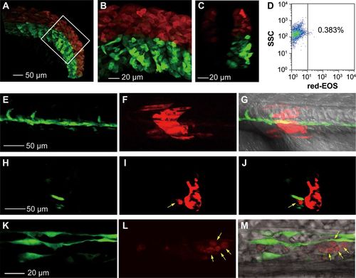
Photoconverted hematopoietic cells are derived from somites but not LPM-derived blood precursors. (A-D) Dorsal somitic cells have a certain hematogenic activity at the 15s stage. The dorsal portion (above the notochord) of all somites in Tg(foxc1b:EOS) embryos was irradiated to avoid flashing the LPM-derived hematopoietic precursors. The posterior trunk region was shown (A) with the boxed area enlarged (B) and an optical cross section shown in C. The blood cells taken from 10 irradiated embryos in group at 30 hpf were analyzed by flow cytometry for red-EOS+ cells (D). (E-M) A downward irradiated single somite generates hematopoietic cells. The eighth somite on one side of a Tg(foxc1b:EOS;fli1a:gfp) embryo at the 20s stage was irradiated from the dorsal downward and the embryo was observed laterally (anterior to the left) by confocal microscopy at the 28s stage. (E-G) Trunk region focusing on the notochord. (H-J) An optical cross section from (E to G) with a photoconverted blood cell (indicated by an arrow) within the vessel. (K-M) An enlarged ICM region. Photoconverted blood cells were indicated by arrows. |
| Genes: | |
|---|---|
| Fish: | |
| Anatomical Terms: | |
| Stage Range: | 14-19 somites to 26+ somites |