
Fig. S6
- ID
- ZDB-FIG-160923-11
- Publication
- Qiu et al., 2016 - Embryonic hematopoiesis in vertebrate somites gives rise to definitive hematopoietic stem cells
- Other Figures
- All Figure Page
- Back to All Figure Page
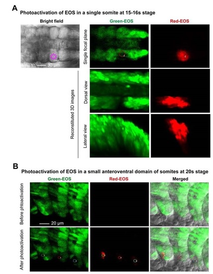
Photoactivation of the reporter EOS in defined embryonic locations. Tg(foxc1b:EOS) transgenic embryos were used throughout. (A) Photoactivation of a single somite at 15-16s stages. The irradiated somite on one side was marked in the left bright-field image (dorsal view). The green EOS in the exposed area started to emit red fluorescence immediately after irradiation (right panel). The embryo was orientated with anterior to the left. Note that fluorescent cells in between the paraxial mesoderm (in the middle panel) should be emigrating somitic cells, either sclerotomal or transdifferentiating hematopoietic cells. (B) A small anteroventral region of three consecutive somites of an embryo at 20s stage was irradiated and red fluorescence was detected by confocal microscopy immediately. |