
Fig. 7
- ID
- ZDB-FIG-140422-45
- Publication
- Holly et al., 2014 - Sfrp1a and Sfrp5 function as positive regulators of Wnt and BMP signaling during early retinal development
- Other Figures
- All Figure Page
- Back to All Figure Page
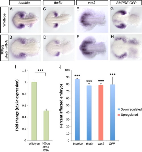
Overexpressing a high dose of sfrp5 mRNA results in loss of dorsal and increase in ventral retina identity. Using in situ hybridization, we analyzed dorsal markers bambia and tbx5a and the ventral marker vax2 at 15 hpf in embryos injected with 100 pg sfrp5 mRNA. Compared to WT, the levels of both tbx5a and bambia expression are significantly reduced in sfrp5 mRNA injected embryos (A–D) while vax2 shows both an increase in expression levels and an expansion of the domain (E–F). We measured BMP signaling using the Tg(BMPRE-AAV.Mlp:eGFP) transgenic strain, which is abbreviated BMPRE:GFP. In situ hybridization for eGFP mRNA in 15 hpf embryos injected with 100 pg sfrp5 mRNA indicates a decrease in BMP signaling compared to wildtype (G–H). Prevalence of change in expression for all probes was quantified and significance was assessed using Fisher′s Exact Test (J,NNN, p<0.0001). qRT-PCR results for tbx5a expression at 15 hpf in sfrp5 mRNA injected embryos indicate a 0.5 fold change in expression (I, NNN, p<0.0001, Student′s t-test). |
| Genes: | |
|---|---|
| Fish: | |
| Anatomical Term: | |
| Stage: | 10-13 somites |
Reprinted from Developmental Biology, 388(2), Holly, V.L., Widen, S.A., Famulski, J.K., and Waskiewicz, A.J., Sfrp1a and Sfrp5 function as positive regulators of Wnt and BMP signaling during early retinal development, 192-204, Copyright (2014) with permission from Elsevier. Full text @ Dev. Biol.