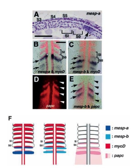
Expression patterns of mesp-a and mesp-b in the presomitic mesoderm. Embryos are oriented with anterior to the top (except for A, in which anterior is to the left). (A) Longitudinal section through a 5-somite stage embryo hybridized with a mesp-a probe. Somites 3, 4 and 5 are labeled as S3, S4 and S5. A newly formed and forming (or the most anterior presumptive) somites are designated as SI and S0, respectively. The mesp-a-positive region (arrow) is located posterior to S0. (B,C) Two-colour staining with myoD (red) and mesp-a (blue in B) or mesp-b (blue in C) at the 10-somite stage. Dorsal views of flat-mounted embryos are shown. Arrows indicate the stripes of mesp genes. (D,E) Two-colour staining with paraxial protocadherin (papc, red) and mesp-b (blue) at the 10-somite stage. Dorsal views under fluorescence (D) and bright-field optics (E) are shown. Arrowheads indicate the anterior borders of papc expression domains and arrows indicate mesp-b expression stripes. Note that both expression domains overlap, sharing the same anterior border. (F) Simplified diagrams illustrating expression patterns of mesp-a, mesp-b, myoD and papc. Bars, 50 μm.
|
