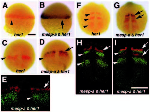
Relationship between mesp-a and her1 expression domains from gastrula to early segmentation period. In all pictures, the probe used is shown at the bottom. Two colour staining with mesp-a and her1 at shield (A,B), 90% epiboly (C-E) and 5-somite (F-I) stages. Lateral views with dorsal to the right (A,B) and dorsal views with anterior to the top (C-I) are shown. Arrowheads indicate her1 expression domains and arrows indicate mesp-a. Hybridized embryos were first processed for her1 (red) staining, photographed, and then processed for mesp-a (blue) staining (A-D,F,G). In E,H and I, hybridized signals were visualized with Fast Red and ELF-97 (see Material and Methods), and the flat-mounted samples were viewed under fluorescent microscopy (mesp-a in red and her1 in green). At the shield stage, the expression domain of mesp-a overlaps with that of her1 (A,B). As gastrulation proceeds, they become completely segregated such that the mesp-a stripes are located more anteriorly (arrow and arrowhead in D and E). During the segmentation period, the anteriormost decaying stripes of the two genes are partially overlapped, while the stripes are seen juxtaposed at the stage when the only one pair of mesp-a strip is observed (F-I). Bars, 100 μm.
|

