
Fig. 5
- ID
- ZDB-FIG-140320-33
- Publication
- Sawada et al., 2000 - Zebrafish Mesp family genes, mesp-a and mesp-b are segmentally expressed in the presomitic mesoderm, and Mesp-b confers the anterior identity to the developing somites
- Other Figures
- All Figure Page
- Back to All Figure Page
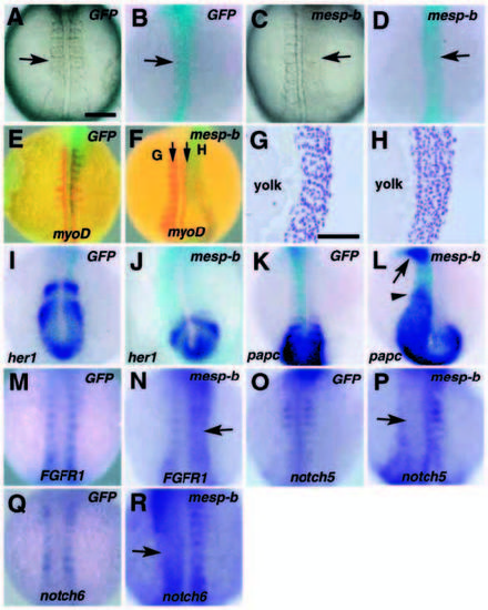
Effect of Mesp-b misexpression on somite formation. Whole-mount samples are viewed dorsally. In all pictures, embryos at the 8- to 12-somite stages are shown and oriented with anterior to the top. The injected RNA is noted in the upper right corner and the probe used is at the bottom. Light blue staining in B,D-F and I-L marks the localization of coinjected β-galactosidase. The samples in M-R were not stained for β-galactosidase activity, which sometimes affects weak in situ staining. The injected embryos showing severe segmentation defects were selected and processed for notch or FGFR1 staining. (A-D) Live embryos injected with GFP (A) or mesp-b (C) and β-galactosidase staining of the same embryos (B,D). Somites fail to form in the mesp-b-injected region (arrow in C,D). (E-H) GFP RNA injection does not affect myoD expression (red staining, E), while segmental myoD expression is either lost or disrupted by the overexpression of mesp-b (F). (G,H) Longitudinal sections of the sample at the levels indicated by the arrows in F. Segmentation is disturbed only in the injected region (H). (I,J) her1 expression in GFP-injected (I) and mesp-b-injected (J) embryos. In the mesp-b-injected embryo (J), light blue staining for b-galactosidase activity is seen on both sides of the embryos. Although her1 expression domains become irregularly shaped, mesp-b injection does not affect the segmental expression of her1. (K,L) papc (blue) expression in GFP-injected (K) and mesp-b-injected (L) embryos. papc loses its segmental expression in the anterior presomitic mesoderm following mesp-b injection. In some cases, papc expression is not down-regulated correctly, resulting in the anteriorly expanded expression (arrowhead in L). Furthermore, expression of papc is sometimes elevated in the head mesenchyme (arrow in L). (M,N) FGFR1 expression in GFP-injected (M) and mesp-b-injected (N) embryos. The expression, which is normally restricted to the presomitic mesoderm and the anterior of formed somites, loses its segmental pattern in the mesp-b-injected region (arrow in N). (O,P) notch5 expression in GFP-injected (N) and mesp-b-injected (O) embryos. The expression, which is normally restricted to the posterior of formed somites (O), is down-regulated in the mesp-b-injected region (arrow in P). (Q, R) notch6 expression in GFP-injected (Q) and mesp-b-injected (R) embryos. The expression, which is normally restricted to the anterior of formed somites, loses its segmental pattern in the mesp-b-injected region (arrow in R). Bars in A and G, 100 and 50 μm respectively. |