
Fig. 4
- ID
- ZDB-FIG-100816-23
- Publication
- Tong et al., 2010 - Zebrafish monosex population reveals female dominance in sex determination and earliest events of gonad differentiation
- Other Figures
- All Figure Page
- Back to All Figure Page
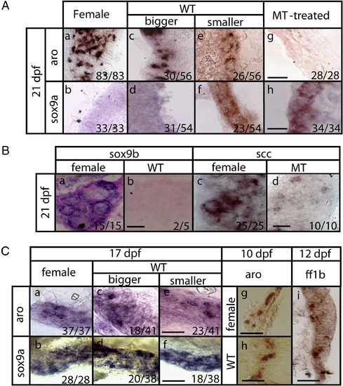
Expression of gonad markers in testes and ovaries at different stages. Expression of sox9a, aro and ff1b in (A) 21-dpf whole-mount (B) 21-dpf cross-sectioned and (C) 17-, 10-, and 12-dfp whole-mount gonads was detected by in situ RNA hybridization. Positive signals are shown in blue or brown color. All sectioned slides were counter-stained with Nuclear Fast Red. All female fish are progeny of super mothers. WT: wildtype fish. Bigger and smaller gonads in (A) and (C) refer to gonad sizes. Numbers at the corner indicate the number of larvae with a typical RNA expression pattern versus the total number of larvae. Scale bars represent 40 μm in (A and Ca–f), 20 μm in (B) and 25 μm in (Cg–i). aro: cyp19a1a, ff1b: nr5a1a, scc: cyp11a1. |
| Genes: | |
|---|---|
| Fish: | |
| Condition: | |
| Anatomical Terms: | |
| Stage Range: | Days 7-13 to Days 21-29 |
Reprinted from Developmental Biology, 344(2), Tong, S.K., Hsu, H.J., and Chung, B.C., Zebrafish monosex population reveals female dominance in sex determination and earliest events of gonad differentiation, 849-856, Copyright (2010) with permission from Elsevier. Full text @ Dev. Biol.