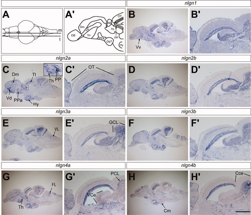
Expression of nlgns in adult brain. A: ISH staining is displayed in saggital sections of adult brain at a medial (m) and a more lateral level (l) as indicated in the dorsal view of adult brain. Medial sections are displayed in B through H and more lateral sections are displayed in B′ through H′. A′: Schematic of the forebrain displays subregions of the telencephalon. B-H: The entire brain. B′-H′: An enlargement of optic tectum, valvula, and corpus cerebelli. The inset in C is an enlargement of the thalamus and periventricular pretectal nucleus. CCe, corpus cerebelli, Cm, corpus mamillare; Dm, medial portion of the dorsal telencephalon; Hy, hypothalamus; OT, optic tectum; PP, periventricular pretectal nucleus; Th, thalamus; Tl, torus longitudinalis; Vd, dorsal portion of the ventral telencephalon; Vv, ventral portion of the ventral telencephalon; GCL, granule cell layer; PCL, Purkinje cell layer; VCe, valvula cerebelli. Scale bar = 750 μm in medial sections and 320 μm in lateral sections.
|
