
Fig. 7
- ID
- ZDB-FIG-090515-38
- Publication
- Martin et al., 1995 - Five Trk receptors in the zebrafish
- Other Figures
- All Figure Page
- Back to All Figure Page
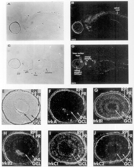
Zebrafish trk expression in the spinal cord, cranial ganglia, and retina. RNA probes labeled with [35S]UTP specific for trk full-length transcripts were used to examine expression 6 days after fertilization, in sagittal sections for trkC1 (A-D) and the retina for the five trk probes (E-J) using in situ hybridization. (B and D) trkC1 dark-field photomicrographs. (A and C) phase photomicrographs of B and D, respectively. (F) trkA, (G) trkB1, (H) trkB2, (I) trkC1, and (J) trkC2 are dark-field photomicrographs. (E) Phase photomicrograph of F (trkA). The positions of retinal pigment epithelium (RPE), photoreceptors (PR), inner nuclear layer (INL), and ganglion cell layer (GCL) are indicated. The lens next to the GCL in F and H appears white but does not correspond to labeling. The scale bar is 50 μm. |
| Genes: | |
|---|---|
| Fish: | |
| Anatomical Terms: | |
| Stage: | Day 6 |
Reprinted from Developmental Biology, 169, Martin, S.C., Marazzi, G., Sandell, J.H., and Heinrich, G., Five Trk receptors in the zebrafish, 745-758, Copyright (1995) with permission from Elsevier. Full text @ Dev. Biol.