
Fig. 6
- ID
- ZDB-FIG-090515-37
- Publication
- Martin et al., 1995 - Five Trk receptors in the zebrafish
- Other Figures
- All Figure Page
- Back to All Figure Page
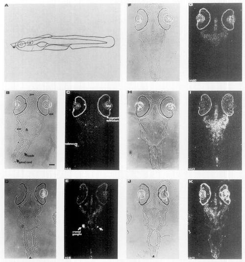
Zebrafish trk genes are expressed in the central and peripheral nervous system. Five RNA probes labeled with [35S]UTP specific for the full-length transcripts of the trk genes were used to examine expression 6 days after fertilization by in situ hybridization. (A) Schematic indicating the most dorsal plane of the sections of the zebrafish embryo. The position of the subsequent sections is indicated by the arrowhead. (C) trkA, (E) trkB1, (G) trkB2, (I) trkC1, and (J) trkC2 are dark-field photomicrographs. B, D, F, H, and J are phase photomicrographs of C, E, G, I, and K, respectively. The m and arrowhead in B marks the cell body of the Mauthner cell. The retinal pigmented epithelium melanocytes and iridiophores contain a chromophore which causes the formation of silver grains in the photographic emulsion and is not hybridization signal. Such a melanocyte (or iridiophore) is labeled with an arrow in C to illustrate this phenomenon. The lens appears white, but this is not hybridization signal, as it appeared the same without hybridization. The scale bar is 50 μm. |
| Genes: | |
|---|---|
| Fish: | |
| Anatomical Terms: | |
| Stage: | Day 6 |
Reprinted from Developmental Biology, 169, Martin, S.C., Marazzi, G., Sandell, J.H., and Heinrich, G., Five Trk receptors in the zebrafish, 745-758, Copyright (1995) with permission from Elsevier. Full text @ Dev. Biol.