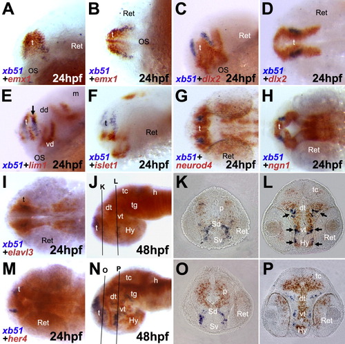
Double in situ hybridization of xb51 and with several molecular markers. Front-lateral (A, C, E, F), dorsal (B, D, G, H, I, M), and lateral views (J, N). Anterior to the left. A-D: At 24 hpf, the expression domain of xb51 is overlapped with that of emx1, a dorsal telencephalon marker (A, B), but not with dlx2, a ventral telencephalon and diencephalon marker (C, D). E-H: The expression domain of xb51 is partly overlapped with that of lim1 in the dorsal telencephalon at 24 hpf (arrow in E), while it is not overlapped with that of islet1 (F), neurod4/zath3 (G), and ngn1 (H). I-L: At 24 hpf, the xb51 expression domain is included in that of elavl3/huC, a pan-neuronal marker (I). At 48 hpf, in cross-section (J-L), the expression domain of xb51 is overlapped with elavl3/huC in the lateral region of the brain (arrows). M-P: The xb51 expression domain is not overlapped with that of her4, a marker for proliferating undifferentiated neural cells. In cross-sections of embryos at 48 hpf (O, P), expression domains for xb51 and her4 show a complementary pattern for each other. Approximate planes of the sections are indicated by lines in J and N. p, pallium; Sd, dorsal subpallium; Sv, ventral subpallium.
|
