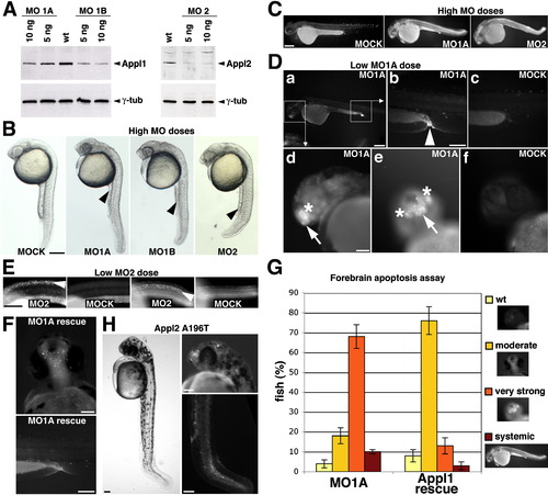
Loss of Appls Causes Apoptosis
(A) Reduction of Appl1 and Appl2 protein levels upon injection of 5 and 10 ng antisense MOs into single-cell stage embryos. Proteins were extracted from 10-somite embryos to illustrate dose-dependent MO-mediated knockdown efficiency. Knockdown efficiency also depends on the stage of extraction (see later extracts, Figure 5A). Two MOs knock down Appl1 (MO1A and MO1B), and one MO knocks down Appl2 (MO2).
(B) Morphological phenotypes of 24 hr zebrafish injected with either injection buffer only (MOCK) or “high” doses of MOs: MO1A (12 ng), MO1B (12 ng), or MO2 (4 ng): reduced yolk extensions and overall sick appearance. The scale bar represents 250 μm.
(C–H) Apoptotic staining by AO (in white) on MOCK-injected, morphant and TILLING zebrafish embryos at 54 hr of development. AO unspecifically accumulates in the yolk.
(C) Wide apoptosis in “high”-dose morphants (MO1A [12 ng] and MO2 [4 ng]). The scale bar represents 250 μm.
(D) Local apoptosis in “low”-dose MO1A morphants (8 ng). (a) Low-dose MO1A embryo, lateral view. Head region magnified in panels (d) and (e) and tails magnified in panel (b) are indicated. (b) Strong apoptosis of the distal pronephric tube. (c) Corresponding region of a MOCK-injected embryo. (d and e) Magnified MO1A head region, lateral (d) and front view (e). Strong apoptosis is observed in forebrain including telencephalon and olfactory placode. (f) Corresponding region of a MOCK-injected embryo (front view). The scale bars represent (a) 250 μm, (b–f) 100 μm.
(E) Local apoptosis in low-dose MO2 morphants (2 ng) is restricted to the neural tube, shown at the level of the yolk extension. Panels to the left, lateral view; panels to the right, top view. The scale bar represents 250 μm.
(F) Rescue of apoptosis in forebrain and pronephros in low-dose MO1A morphants (8 ng) by coinjection of 75 pg appl1 mRNA that lacks the MO1A binding site. The scale bars represent 100 μm.
(G) Forebrain apoptosis assay: Quantitative evaluation of apoptosis in Appl1 knockdown (MO1A: 8 ng MO1A) and rescued zebrafish embryos (Appl1 rescue: 8 ng MO1A + 75 pg appl1 mRNA). Embryos were assigned to one of four cell death categories: WT, moderate, very strong forebrain, and systemic apoptosis (representative images in legend). Total amount of embryos scored: n MO1A = 532; n Appl1 rescue = 598. Error bars indicate SD between experiments. Note that apoptosis is significantly rescued in the majority of embryos.
(H) APPL2A196T mutant fish isolated by TILLING are characterized by reduced yolk extension, kinked tails, apoptosis in the neural tube, and olfactory placodes, resembling appl2 morphants (left panel). The scale bars represent 100 μm.
|
