
FIGURE
Fig. S2
- ID
- ZDB-FIG-080508-11
- Publication
- Schenck et al., 2008 - The endosomal protein Appl1 mediates Akt substrate specificity and cell survival in vertebrate development
- Other Figures
- All Figure Page
- Back to All Figure Page
Fig. S2
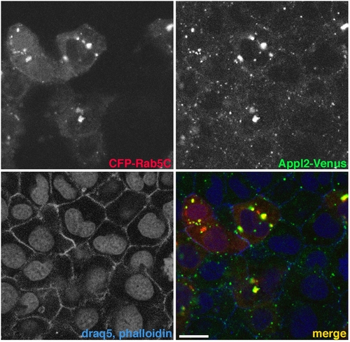
Appl2 localization and recruitment to Rab5C-labeled endosomes. |
Expression Data
Expression Detail
Antibody Labeling
Phenotype Data
Phenotype Detail
Acknowledgments
This image is the copyrighted work of the attributed author or publisher, and
ZFIN has permission only to display this image to its users.
Additional permissions should be obtained from the applicable author or publisher of the image.
Reprinted from Cell, 133(3), Schenck, A., Goto-Silva, L., Collinet, C., Rhinn, M., Giner, A., Habermann, B., Brand, M., and Zerial, M., The endosomal protein Appl1 mediates Akt substrate specificity and cell survival in vertebrate development, 486-497, Copyright (2008) with permission from Elsevier. Full text @ Cell