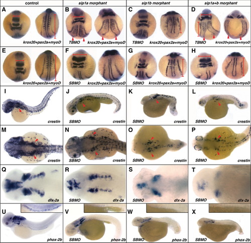
Effect of sip1a and sip1b translation and splice blocking morpholino antisense oligonucleotide injection on axial patterning, and neural crest specification and migration. (A, E, I, M, Q, U) Wild-type control, (B) sip1a TBMO morphant, (F, J, N, R, V) sip1a SBMO morphant, (C) sip1b TBMO morphant, (G, K, O, S) sip1b SBMO morphant embryos, (D, H, L, P, T) sip1a sip1b double morphant embryos. A-H: Dorsal views of 3-somite stage embryos that have been hybridized with a krox20, pax2a, and myoD riboprobes mix. I-L: Lateral views of 36hpf embryos that have been hybridized with riboprobes for crestin. Lateral (M-P) and dorsal (Q-T) views of 48hpf embryos that have been hybridized with riboprobes for dlx-2a. U-X: Lateral views of 55hpf embryos that have been hybridized with riboprobes for phox2b. Insets in U-X are close-ups of the intestine of these embryos. Red arrowheads (B-D) point to convergence defects in the midline. Red horizontal bars (A, B, D, E, F, H) highlight flattening of the morphant embryos in the hindbrain region, as compared to the control. Red vertical bars (A, C, D, E, G, H) highlight shorter axis in morphant embryos, as compared to control. Red arrowheads (I-P) indicate the vagal/post-otic neural crest. Anterior is to the left.
|
