
Fig. 4
- ID
- ZDB-FIG-070117-3
- Publication
- Krens et al., 2006 - Characterization and expression patterns of the MAPK family in zebrafish
- Other Figures
- All Figure Page
- Back to All Figure Page
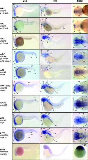
Expression patterns of the zebrafish MAPK genes by in situ hybridization. The first two columns show 24 and 48 hpf old zebrafish, lateral view, anterior to left, dorsal to top. The third column shows a dorsal view of the head region, anterior to left, at 48 hpf. All MAPK genes are indicated with all known names. MAPKs erk3 and erk4 are indicated with an astrix (*) because of their alternative dual phosphorylation site (SEG). All MAPK were expressed and showed distinct expression patterns at 24 and 48 hpf, but p38b. Therefore the dorsal head-image is replaced for an of the expression patterns at high stage (∼3.5 hpf), where p38b was expressed in the whole animal pole. ce, cerebellum; di, diencephalon; gu, gut; hb, hindbrain; hy, hypothalamus; nt, notochord, op, olfactory pit / placode; pf, pectoral fin; pnd, pronephric ducts; pr, proctodaeum re, retina; sb, somite boundary; tb, tail bud; tc, tectum; te, telencephalon; tg, tegmentum; tm, tail muscle. |
Reprinted from Gene expression patterns : GEP, 6(8), Krens, S.F., He, S., Spaink, H.P., and Snaar-Jagalska, B.E., Characterization and expression patterns of the MAPK family in zebrafish, 1019-1026, Copyright (2006) with permission from Elsevier. Full text @ Gene Expr. Patterns