
- Title
-
Antagonistic interactions safeguard mitotic propagation of genetic and epigenetic information in zebrafish
- Authors
- Lawir, D.F., Soza-Ried, C., Iwanami, N., Siamishi, I., Bylund, G.O., O Meara, C., Sikora, K., Kanzler, B., Johansson, E., Schorpp, M., Cauchy, P., Boehm, T.
- Source
- Full text @ Commun Biol
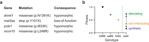
Study design. |
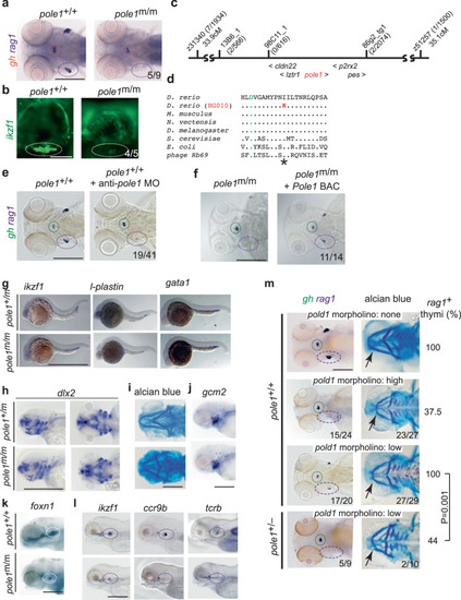
Characterization of a zebrafish |
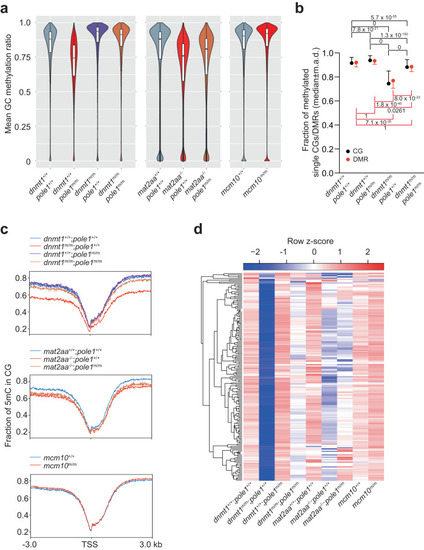
Global changes in DNA methylation patterns in single and double mutant zebrafish. |
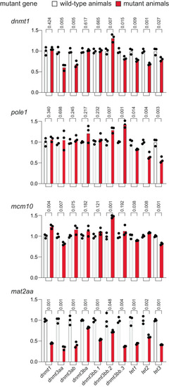
Expression analysis of genes associated with the DNA methylation process. qPCR analysis was performed on 5 dpf embryos of wild-type and mutants ( |
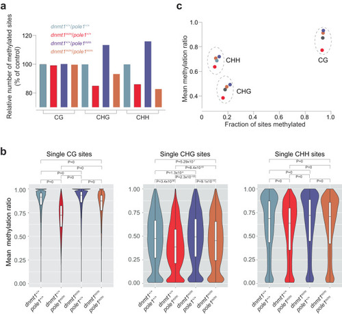
Alterations in non-CG methylation patterns in |
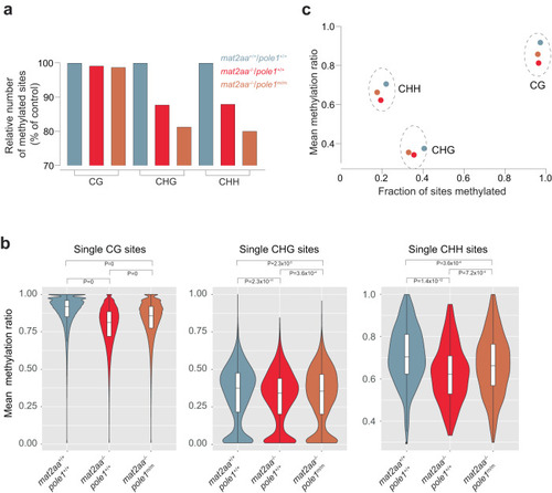
Alterations in non-CG methylation patterns in |

Transcriptional landscapes in |
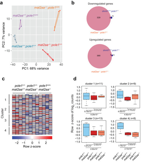
Transcriptional landscapes in |
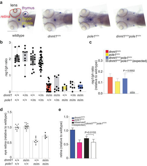
Epistasis analysis of |
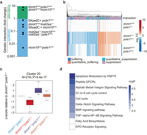
Genetic interaction analysis. |