
- Title
-
nkx2.3 is responsible for posterior pharyngeal cartilage formation by inhibiting Fgf signaling
- Authors
- Yang, S., Xu, X., Yin, Z., Liu, Y., Wang, H., Guo, J., Wang, F., Bao, Y., Zhang, T., Sun, S.
- Source
- Full text @ Heliyon
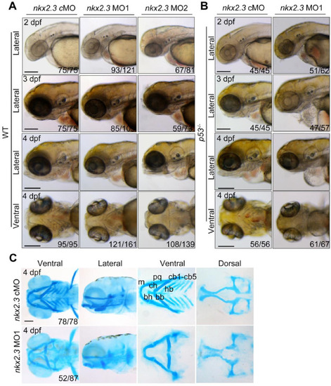
Malformation of pharyngeal cartilages in nkx2.3 morphants. (A) Morphology of WT embryos injected with nkx2.3 MOs at indicated stages. Scale bars, 200 μm. (B) Morphology of p53 mutant embryos injected with nkx2.3 MOs at 4 dpf. Scale bars, 200 μm. (C) Detection of craniofacial cartilages formation using Alcian blue staining. The cartilages were shown anterior to the left and included the ceratobranchial (cb), ceratohyal (ch), Meckel's cartilage (m), basibranchial (bb), basihyal (bh), hypobranchial(hb), and palatoquadrate (pq) cartilages. Scale bars, 100 μm. PHENOTYPE:
|
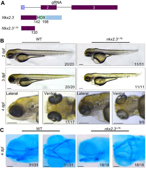
Malformation of pharyngeal cartilages in nkx2.3 mutants. (A) Knockout of nkx2.3 by CRISPR-Cas9 system. The nkx2.3△76 allele was identified by the targeting of the second exon of the nkx2.3 locus (top panel), which carried a 76-bp deletion resulting in a premature stop codon. The truncated protein in nkx2.3△76 mutants only has 130 amino acids (bottom panel). (B) Morphology of nkx2.3 mutant embryos at indicated stages. Scale bars, 200 μm. (C) Photomicrographs of the cartilages stained with Alcian blue. The cartilages were shown anterior to the left and included the ceratobranchial (cb), ceratohyal (ch), Meckel's cartilage (m), and palatoquadrate (pq) cartilages. Scale bars, 100 μm. |
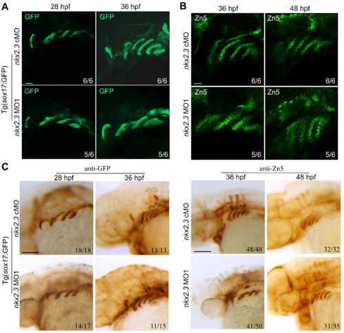
Pharyngeal pouches are normally developed in nkx2.3 morphants. (A) Embryos were immunostained with the anti-GFP antibody at the indicated stages. Scale bars, 20 μm. (B) Embryos were immunostained with the anti-Zn5 antibody at the indicated stages. Scale bars, 20 μm. (C) Embryos were immunostained with the anti-Zn5 antibody at the indicated stages. Scale bars, 100 μm. Images are shown with a lateral view and with the anterior to the left. |
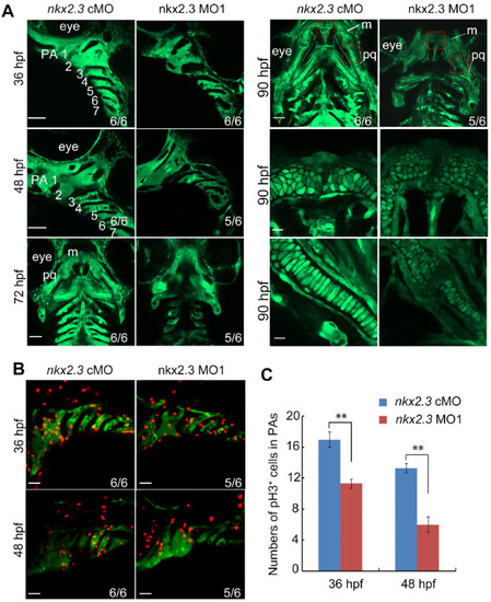
Proliferation of CNCCs is compromised in nkx2.3 morphants. (A) Time-lapse imaging of nkx2.3 morphants and control embryos in Tg(fli1-GFP) from 36 to 90hpf. The bottom right is an enlarged view of the red box in the top right figure at 90 hpf. Scale bars: 20 μm. (B) Immunostaining of embryos in the background of Tg(fli1:GFP) with pH3 at indicated stages. Scale bars: 20 μm. (C) The number of pH3 and GFP double-positive cells in the pharyngeal arches. Data are represented as mean ± SD. **P < 0.01. EXPRESSION / LABELING:
PHENOTYPE:
|
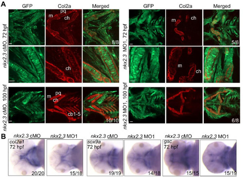
Posterior arch skeletogenic CNCCs do not differentiate in nkx2.3 morphants. (A) Detection of Col2a proteins in the pharyngeal arches at indicated stages. Embryos of Tg(fli1:EGFP) injected with nkx2.3 MOs were co-immunostained with anti-GFP (green) and anti-Col2a (red) antibodies. The palatoquadrate (pq), ethmoid plate (ep) and ceratohyal (ch) cartilages were shown with anterior to the left. Scale bars, 20 μm. (B) The expression level of marker genes of CNCCs’ differentiation was detected by WISH at 72 hpf. EXPRESSION / LABELING:
PHENOTYPE:
|
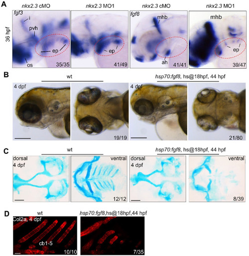
Knockdown of nkx2.3 increased Fgf signal and fgf8 overexpression mimic the phenotype of nkx2.3 morphants. (A) Expression pattern of fgf3 and fgf8 in nkx2.3 morphants detected by ISH at 36 hpf. i, isthmus; os, optic stalk; pvh, posterior-ventral hypothalamus; ep, endodermal pouches; mhb, midbrain-hindbrain boundary; ah, adenohypophysis. (B) Morphology of embryos with overexpressed fgf8. Scale bars: 200 μm. (C) Alcian blue staining of embryos in which fgf8 was overexpressed. Scale bars: 100 μm. (D) Immunostaining of Col2a proteins in fgf8 transgenic embryos at 4 dpf. Scale bars: 10 μm. |
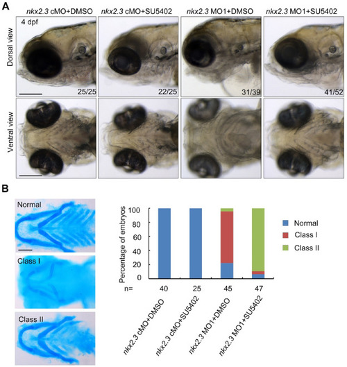
Fgf signal inhibition can partially correct the malformation of the posterior cartilages in nkx2.3 morphants. (A) Embryo morphology following SU5402 treatment on nkx2.3 morphants. We tracked the embryos' growth and took pictures of them at 4 dpf. Embryos treated with 2.5 μM SU5402 between 24 and 48 hpf after receiving an injection of nkx2.3 cMO or nkx2.3 MO1 are shown in the representative images. Scale bars: 200 μm. (B) Alcian blue staining of nkx2.3 morphants treated with SU5402 and the statistic results. At 4 dpf, the embryos of nkx2.3 morphants and controls that had been treated with of 2.5 μM SU5402 were collected and stained with Alcian blue. Representative images of the various pharyngeal cartilage classes were shown in the left panel. Scale bars: 100 μm. The bar graph on the right displays the proportion of each type of embryo seen in the left panel in each treatment group. PHENOTYPE:
|