
- Title
-
Encapsulation of the septal cell wall protects Streptococcus pneumoniae from its major peptidoglycan hydrolase and host defenses
- Authors
- Figueiredo, J., Henriques, M.X., Catal�o, M.J., Pinheiro, S., Narciso, A.R., Mesquita, F., Saraiva, B.M., Carido, M., Cabanes, D., Pinho, M.G., Filipe, S.R.
- Source
- Full text @ PLoS Pathog.
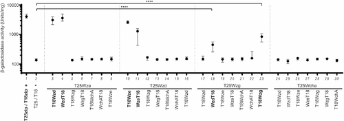
Fig 1. Wzg, a putative CPS-cell wall ligase, interacts with Wzd and with itself. Interactions between Wze, Wzd, Wzg and WchA were tested using Bacterial Two-Hybrid E. coli system [31]. ?-galactosidase activity of cells expressing putative interaction partners was measured in cell extracts in at least three independent replicates. Black circles indicate median values and brackets show the 25% and 75% percentiles. Positive control (+): E. coli expressing T18 and T25 fragments linked to leucine zipper domains (zip) that can dimerize; Negative control (-): E. coli expressing untagged T18 and T25 fragments. Interactions were detected between T25-Wzg/Wzd-T18 and T25-Wzg/T18-Wzg. ****P???0.0001. As previously described [28] combinations of plasmids expressing Wzd and Wze indicate that these proteins interact. |
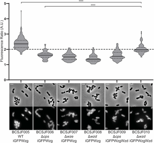
Fig 2. Septal localization of Wzg is dependent on the expression of Wzd/Wze. Graph shows the ratio of iGFP-Wzg fluorescence measured at the septum versus the peripheral wall in the S. pneumoniae wild-type encapsulated strain (BCSJF005, n = 194), in the cps null mutant (BCSJF006, n = 121), in the wze null mutant (BCSJF007, n = 106), in the wzd null mutant (BCSJF008, n = 107) and in the cps and wzd null mutants expressing Wzd from a constitutive promoter (BCSJF009, n = 112, and BCSJF010, n = 109, respectively). Enrichment of Wzg at the septum is only observed when Wzd is expressed and is localized at the division septum. Solid lines indicate median, and dashed lines indicate 25% and 75% percentiles. Representative phase contrast and fluorescence microscopy images of each strain are shown below the graph. ****P???0.0001. Scale bar, 2 ?m. |
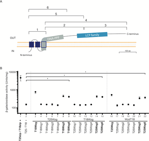
Fig 3. The transmembrane and DNA-PPF domains of Wzg are required for its self-interaction and interaction with Wzd. A) Scheme indicating the 6 different regions of Wzg tested for interaction with Wzd by BTH. B) Interactions between Wzg and the Wzg1-6 constructs were analyzed by BTH. Black circles indicate median values and brackets show the 25% and 75% percentiles of ?-galactosidase activity measured in cell extracts of at least three independent replicates. Positive control (+): E. coli expressing T18 and T25 fragments linked to leucine zipper domains that can dimerize; Negative control (-): E. coli expressing untagged T18 and T25 fragments. *P???0.05. Only the Wzg5 and Wzg6 constructs, which include the three transmembrane domains and the DNA-PPF domain, interact with full-length Wzg and with Wzd. |
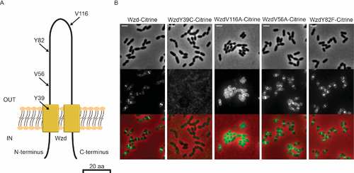
Fig 4. Point mutations in Wzd affect its localization. A) Scheme of Wzd protein indicating the localization, in the extracellular loop, of four mutations that were associated with a mucoid colony phenotype [19]. B) Localization of Citrine fluorescent derivatives of Wzd single-residue mutants expressed in a wzd null mutant strain. Strain BCSMH022, expressing Wzd-Citrine was used as control. V56A and Y82F mutations did not interfere with the ability of Wzd to localize at the division septum upon interaction with Wze. Representative phase contrast (top panels, for visualization of bacteria), fluorescence microscopy (middle panels, for detection of the capsule associated with the bacterial cell surface) and overlay (bottom panels) images of each strain are shown. Scale bar, 2 ?m. |
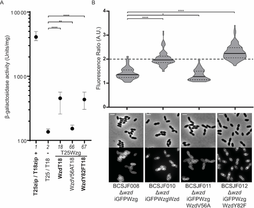
Fig 5. The valine residue at position 56 of Wzd is required for its interaction with Wzg, and for the recruitment of Wzg to the division septum. A) Interaction between Wzg and Wzd, WzdV56A and WzdY82F point mutants was tested by BTH. Black circles indicate median values and brackets show the 25% and 75% percentiles of ?-galactosidase activity measured in cell extracts of at least three independent replicates. Positive control (+): E. coli expressing T18 and T25 fragments linked to leucine zipper domains (zip) that can dimerize; Negative control (-): E. coli expressing untagged T18 and T25 fragments. WzdV56A-T18 lost the ability to interact with T25-Wzg. B) Graph shows the ratio of iGFP-Wzg fluorescence measured at the septum versus the peripheral wall in the S. pneumoniae wzd null mutant strain (BCSJF008, n = 107) and in the wzd null mutant strain expressing Wzd or Wzd point mutants from a constitutive promoter (non-mutated Wzd strain BCSJF010, n = 109, mutant WzdV56A strain BCSJF011, n = 106, and mutant WzdY82F strain BCSJF012, n = 128). WzdY82F, but not WzdV56A, can recruit Wzg to the division septum of pneumococcal bacteria. *P???0.05, **P???0.01, ****P???0.0001. |
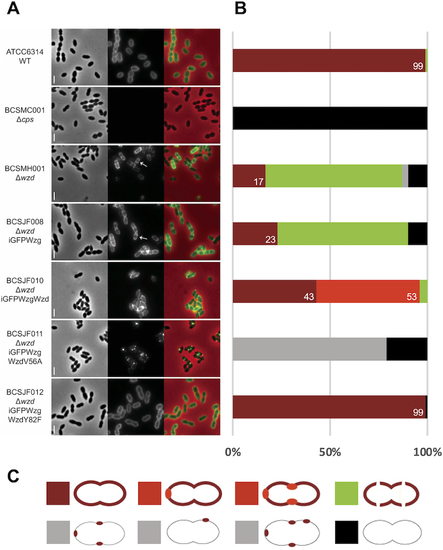
Fig 6. Expression of WzdV56A impairs the Wzg ability to produce a pneumococcal cell surface fully surrounded by capsule. A) Immunofluorescence microscopy images using a serotype-14 specific serum to detect the presence of the capsular polysaccharide at the cell surface. Wild-type encapsulated ATCC6314 (n = 115) expressed capsule all over the surface, while wzd null mutant strain BCSMH001 lacked capsule at midcell (n = 115). Cells of wzd null mutant strain were transformed with plasmids expressing (i) iGFP-Wzg alone (BCSJF008, n = 108), resulting in cells where the capsule is absent from the division septum; (ii) iGFP-Wzg and Wzd (BCSJF010, n = 104), or with WzdY82F (BCSJF012, n = 117), resulting in cells with homogeneous distribution of CPS at their surface or (iii) iGFP-Wzg and WzdV56A (strain BCSJF011, n = 105), resulting in cells where CPS accumulated in spots and was absent from most of the bacterial cell surface. Representative phase contrast (top panels, for visualization of bacteria), fluorescence microscopy (middle panels, for detection of the capsule associated with the bacterial cell surface) and overlay (bottom panels) images of each strain are shown. Arrows highlight bacteria that lack CPS at midcell. Scale bar, 2 ?m. B) Graph shows the percentage of cells, for each strain, with different CPS patterns (grouped in 5 different classes). Numbers represent the percentage of cells fully covered with CPS. C) Classes of CPS patterns: cells fully covered with homogenous CPS (dark red); cells fully covered with CPS whose staining is heterogenous and has brighter regions (light red); cells partially covered with interruptions at the division septum (green); cells with CPS only in spots in particular regions (grey) or lacking CPS (black). |
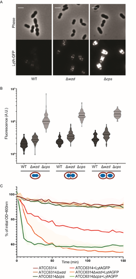
Fig 7. Absence of capsule at the division septum results in increased exposure of bacteria cell wall and susceptibility to lysis. A) Epifluorescence microscopy images of wild-type ATCC6314 strain (WT), its capsule null mutant (BCSMC001; ?cps) and the wzd null mutant (BCSMH001; ?wzd) incubated with LytA-GFP. Scale bar, 2 ?m. B) Quantification of cell bound LytA-GFP fluorescence. Bacteria were grouped in three different classes (n> 50), depending on their cell cycle stage: (I) recently divided cells; (II) cells initiating division as seen from invagination of cell surface; (III) cells at the final steps of division, with deep invagination at division septum. Lack of capsule (at mid-cell or over the entire cell wall) results in bacteria that bind more LytA-GFP than the parental encapsulated bacteria. C) Lysis of boiled bacterial cells of wild-type ATCC6314, unencapsulated (ATCC6314?cps) and wzd null mutant (ATCC6314?wzd) strains was assessed by the decrease in OD 600 nm in the absence (n = 3) or presence (n = 7) of LytA-GFP. Lysis curves show that the lack of capsule at particular sub-cellular sites of the bacterial cell surface increase bacteria susceptibility to lysis. Solid lines represent the curve obtained with the median values for each timepoint. Shaded areas associated with each solid line represent the interquartile range. |
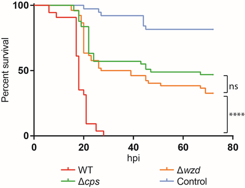
Fig 8. Full encapsulation of the cells is necessary for virulence. A) Survival graph of zebrafish embryos injected, three days post-fertilization, with wild-type ATCC6314 (WT), unencapsulated BCSMC001 (?cps), wzd null mutant BCSMH001 (?wzd) and medium used to resuspend bacteria (Control). The wzd null mutant was severely impaired in virulence, similarly to the unencapsulated BCSMC001 strain. The assay was performed with at least 50 embryos for each condition that were injected in four different days and the obtained survival curves, except those obtained with the cps and wzd mutants, were significantly different (p<0.0001). (hpi) hours post infection. PHENOTYPE:
|