
|
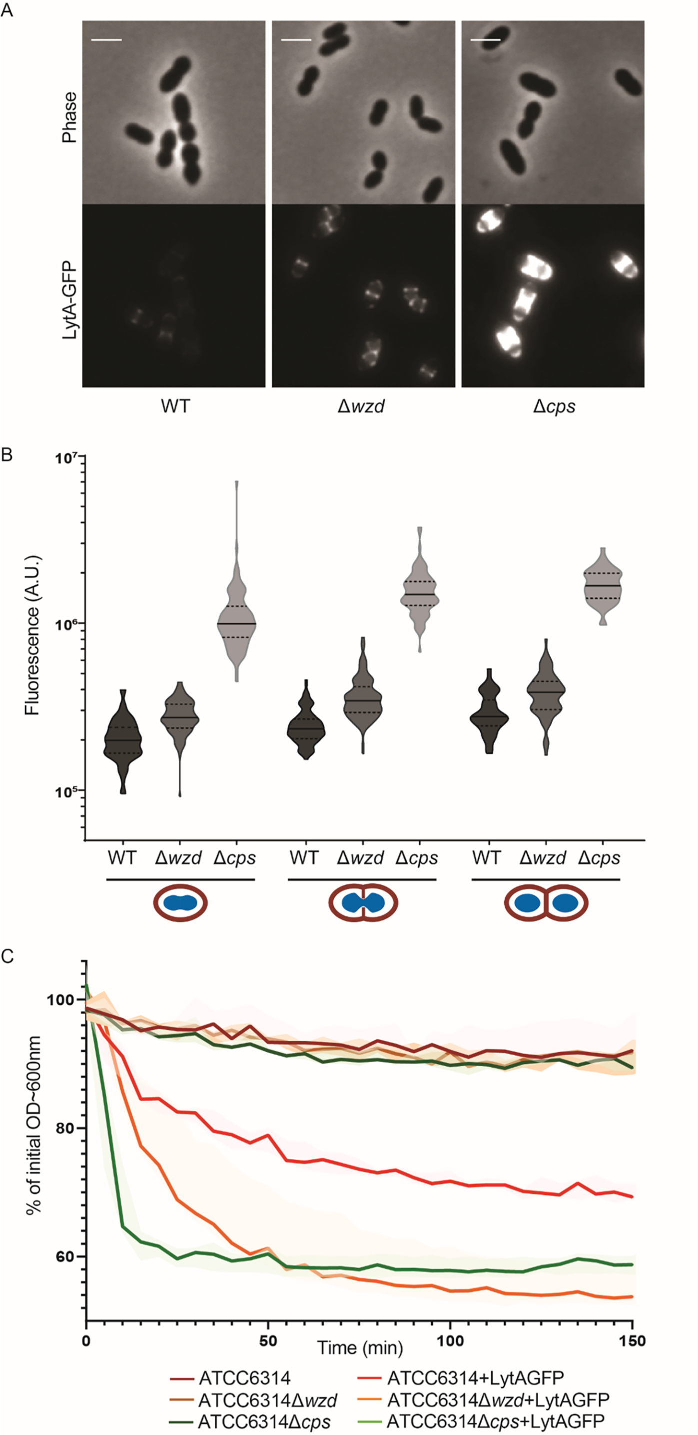
Fig. 7 Fig 7. Absence of capsule at the division septum results in increased exposure of bacteria cell wall and susceptibility to lysis.
A) Epifluorescence microscopy images of wild-type ATCC6314 strain (WT), its capsule null mutant (BCSMC001; Δcps) and the wzd null mutant (BCSMH001; Δwzd) incubated with LytA-GFP. Scale bar, 2 μm. B) Quantification of cell bound LytA-GFP fluorescence. Bacteria were grouped in three different classes (n> 50), depending on their cell cycle stage: (I) recently divided cells; (II) cells initiating division as seen from invagination of cell surface; (III) cells at the final steps of division, with deep invagination at division septum. Lack of capsule (at mid-cell or over the entire cell wall) results in bacteria that bind more LytA-GFP than the parental encapsulated bacteria. C) Lysis of boiled bacterial cells of wild-type ATCC6314, unencapsulated (ATCC6314Δcps) and wzd null mutant (ATCC6314Δwzd) strains was assessed by the decrease in OD 600 nm in the absence (n = 3) or presence (n = 7) of LytA-GFP. Lysis curves show that the lack of capsule at particular sub-cellular sites of the bacterial cell surface increase bacteria susceptibility to lysis. Solid lines represent the curve obtained with the median values for each timepoint. Shaded areas associated with each solid line represent the interquartile range.