
|
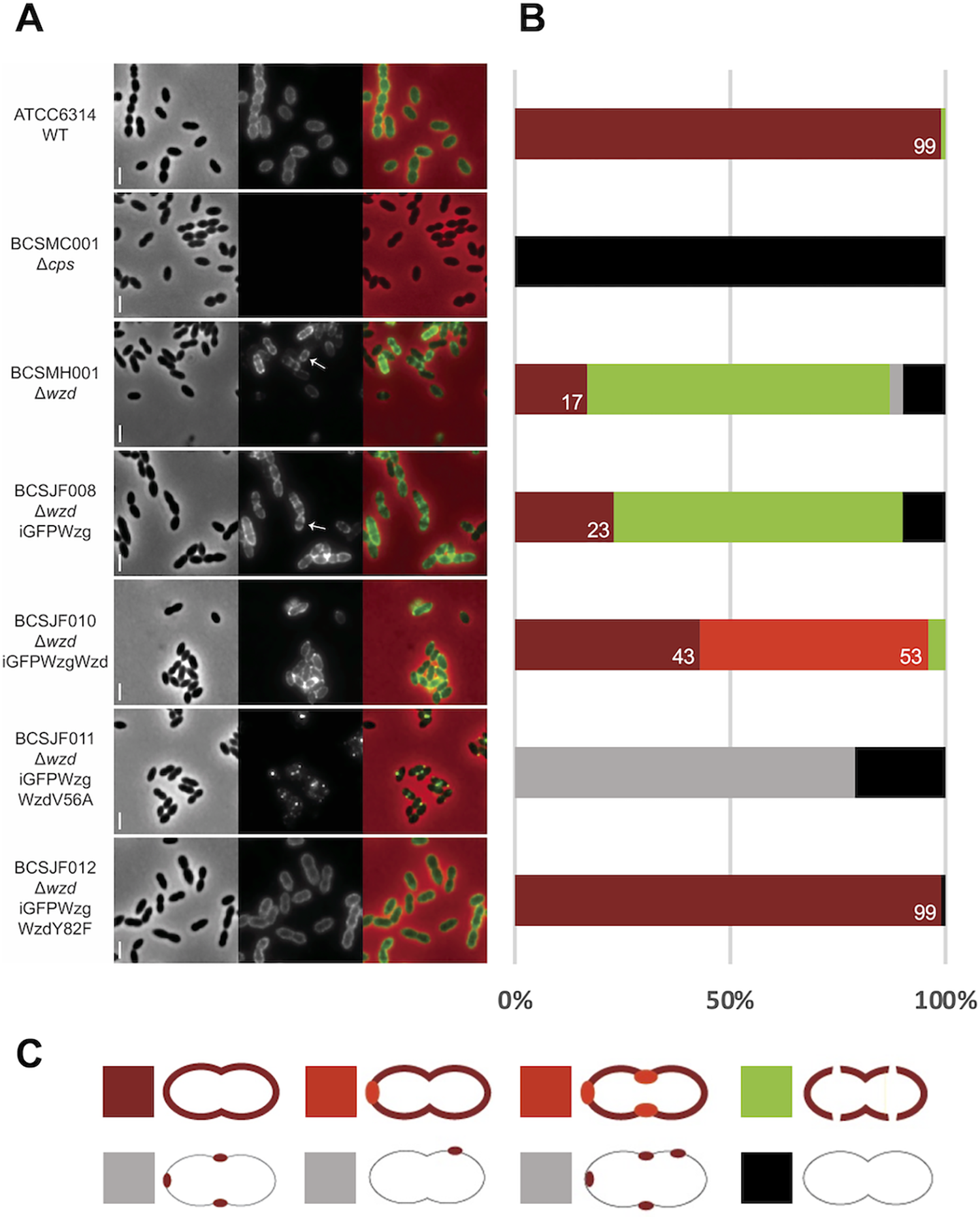
Fig. 6 Fig 6. Expression of WzdV56A impairs the Wzg ability to produce a pneumococcal cell surface fully surrounded by capsule. A) Immunofluorescence microscopy images using a serotype-14 specific serum to detect the presence of the capsular polysaccharide at the cell surface. Wild-type encapsulated ATCC6314 (n = 115) expressed capsule all over the surface, while wzd null mutant strain BCSMH001 lacked capsule at midcell (n = 115). Cells of wzd null mutant strain were transformed with plasmids expressing (i) iGFP-Wzg alone (BCSJF008, n = 108), resulting in cells where the capsule is absent from the division septum; (ii) iGFP-Wzg and Wzd (BCSJF010, n = 104), or with WzdY82F (BCSJF012, n = 117), resulting in cells with homogeneous distribution of CPS at their surface or (iii) iGFP-Wzg and WzdV56A (strain BCSJF011, n = 105), resulting in cells where CPS accumulated in spots and was absent from most of the bacterial cell surface. Representative phase contrast (top panels, for visualization of bacteria), fluorescence microscopy (middle panels, for detection of the capsule associated with the bacterial cell surface) and overlay (bottom panels) images of each strain are shown. Arrows highlight bacteria that lack CPS at midcell. Scale bar, 2 μm. B) Graph shows the percentage of cells, for each strain, with different CPS patterns (grouped in 5 different classes). Numbers represent the percentage of cells fully covered with CPS. C) Classes of CPS patterns: cells fully covered with homogenous CPS (dark red); cells fully covered with CPS whose staining is heterogenous and has brighter regions (light red); cells partially covered with interruptions at the division septum (green); cells with CPS only in spots in particular regions (grey) or lacking CPS (black).