
- Title
-
Heteronemin Suppresses Lymphangiogenesis through ARF-1 and MMP-9/VE-Cadherin/Vimentin
- Authors
- Chen, H.L., Su, Y.C., Chen, H.C., Su, J.H., Wu, C.Y., Wang, S.W., Lin, I.P., Chen, C.Y., Lee, C.H.
- Source
- Full text @ Biomedicines
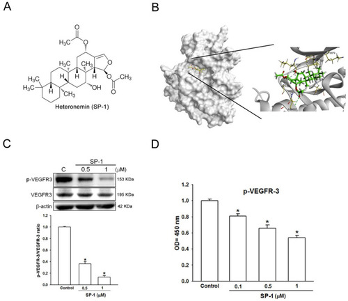
Heteronemin is a VEGFR-3 binding compound in human lymphatic endothelial cells. (A) The structure of heteronemin (indicated as SP-1). (B) Docking models of SP-1-targeted VEGFR-3. The structure of VEGFR-3 was downloaded from PDB (accession: 4BSJ_A) and represented as gray. SP-1, drawn by ChemDraw Ultra 9.0, rendered in the representation of green stick. Close-up of SP-1 docking site (best energy mode) was prepared using Discovery Studio 4.1. (C) Human lymphatic endothelial cells (LECs) were treated with SP-1. Then, the expression of VEGFR-3 and phospho-VEGFR-3 (p-VEGFR-3) was determined by Western blot analysis. The quantitative densitometry of the relative levels of VEGFR-3 and phospho-VEGFR-3 was measured by Image-Pro Plus. (D) Cells were stimulated with SP-1; the phospho-VEGFR-3 protein expression in cell lysate was measured by ELISA. A representation experiment is shown as the mean ± S.D. for three wells (* p < 0.05). Similar results were observed from three independent experiments. |
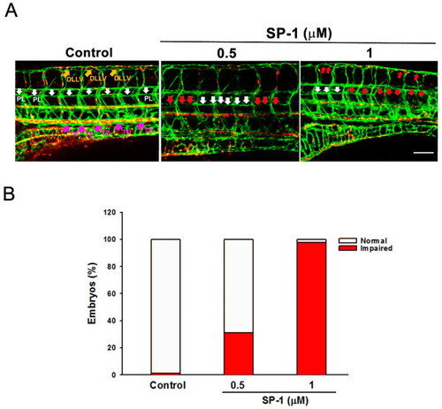
Heteronemin blocks thoracic duct development in zebrafish model. Transgenic (fli1:EGFP;gata1:DsRed) zebrafish embryos were treated with the indicated concentrations of heteronemin (represented as SP-1), and then thoracic duct length was analyzed via fluorescence microscopy. DLLV indicated as dorsal longitudinal lymphatic vessel (orange arrows); PL, parachordal LEC (white arrows) and TD, thoracic duct (pink arrows). (A) Representative pictures of thoracic duct of zebrafish treated with indicated concentration of SP-1 (bar = 100 μm). (B) Quantification of the defective formation of thoracic duct at 5 dpf determined by the percentages of embryos. A total of 50 embryos were analyzed in each experimental condition. |
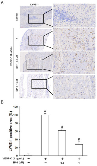
Heteronemin reduces the VEGF-C-stimulated lymphangiogenesis in mouse ear collagen sponges. Gelatin sponges were saturated with medium containing VEGF-C (1 μg/mL; positive control) with or without indicated concentration of heteronemin (represented as SP-1). Sponges were implanted, for 3 weeks, between the two layers of skin in mice ears. (A) Lymphatic vasculatures in sponge sections were detected by LYVE-1 immunostainings (bars = 100 and 70 μm higher magnification). (B) Quantification of the densities of lymphatic vessels (LYVE-1 positive area). Data are expressed as means ± SEM of five mice. * p < 0.05 versus medium sponges; # p < 0.05 versus VEGF-C-stimulated sponges. |
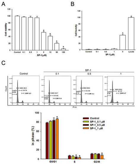
Heteronemin inhibited cell growth and arrested the distribution of cell cycle in human lymphatic endothelial cells. (A) LECs were incubated with various doses (0–100 μM) of heteronemin (indicated as SP-1) in a 96-well plate for 24 h. Cell viability was determined by MTT assay after treatment. The maximal non-toxic dose was chosen for further experiments. (B) Cells were treated with SP-1 for 8 h; then, the cytotoxicity was determined using LDH assay. (C) Cells were treated with various concentrations of SP-1 for 24 h and were then analyzed using flow cytometry. Histograms represent the percentage of cells in each cell cycle phase. The graph corresponds to the distribution of cell subpopulation percentages expressed as means ± SEM of five independent assays. * p < 0.05 compared with solvent control (0.01% DMSO). |
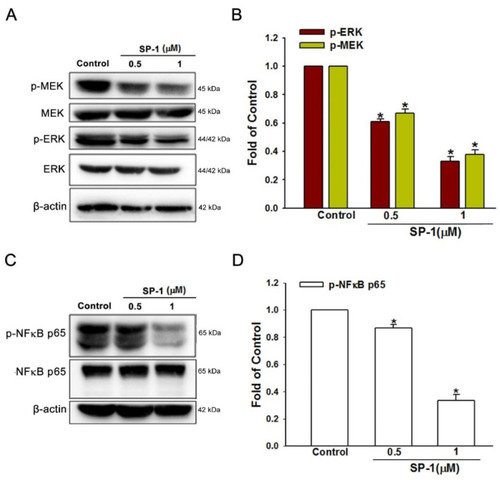
Heteronemin inhibited mitogen-activated protein kinase and transcription factors in human lymphatic endothelial cells. Cells were treated with the indicated concentrations of heteronemin (represented as SP-1); subsequently, the indicated (A) MEK/ERK and (C) p65 phosphorylated and total proteins were determined by Western blot analysis. The quantitative densitometry of phosphorylated (B) MEK/ERK and (D) p65 protein was performed with Image-Pro Plus. Data are expressed as mean ± SEM of five independent experiments. * p < 0.05, compared with the control group. |
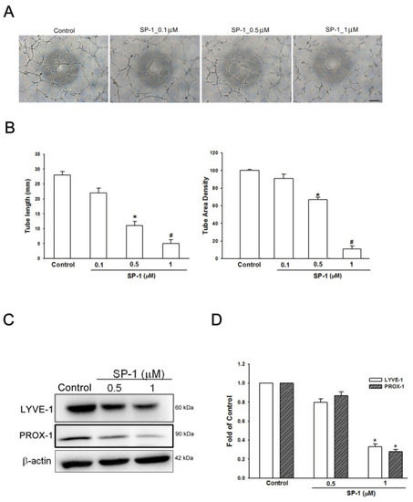
Heteronemin suppressed the tube formation, migration and phosphorylation of VEGF-C/VEGFR-3/LYVE-1 in human lymphatic endothelial cells. ( |
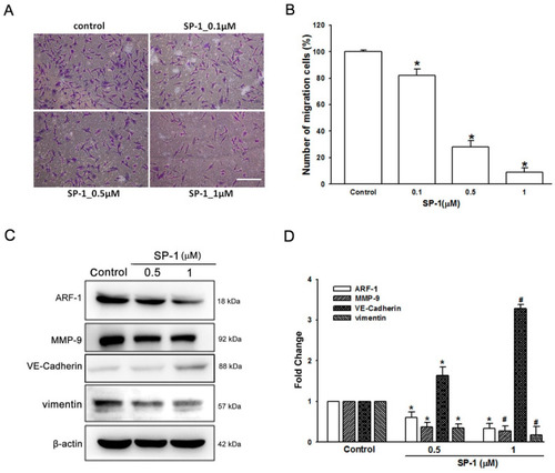
Heteronemin suppressed the migration and endothelial-to-mesenchymal transition of human lymphatic endothelial cells. Cells were treated with the indicated concentrations of heteronemin (represented as SP-1). (A) Cell migration was examined by Transwell assays (scale bar=100 µm; magnification ×100). (B) The quantification of migration was performed using Image-J to validate the anti-lymphangiogenic function of SP-1. (C) LECs were treated with SP-1. Then, the expression of ARF-1 and endothelial-mesenchymal transition-related proteins (MMP-9, VE-cadherin and vimentin) was determined by Western blot analysis. (D) The quantitative densitometry of the relative levels of ARF-1, MMP-9, VE-cadherin and vimentin was measured by Image-Pro Plus. Data are expressed as the mean ± SEM of at least three independent experiments. * p < 0.05, # p < 0.01 compared with the control group. |
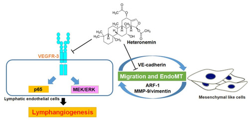
Schema of heteronemin-induced anti-lymphangiogenic mechanism in human lymphatic endothelial cells. This study reveals heteronemin as a promising anti-lymphangiogenic agent. Heteronemin may possess anti-lymphangiogenesis effect by reducing VEGFR-3 and its downstream NF-κB p65 and MEK/ERK signaling pathways and ARF-1, as well as the induction of VE-cadherin and the reduction in MMP-9 and vimentin, which is associated with EndoMT, in human lymphatic endothelial cells. T-shaped lines indicate inhibition. |