
|
Figure 1
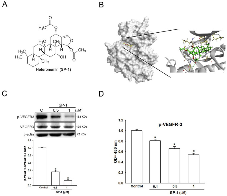
Heteronemin is a VEGFR-3 binding compound in human lymphatic endothelial cells. (A) The structure of heteronemin (indicated as SP-1). (B) Docking models of SP-1-targeted VEGFR-3. The structure of VEGFR-3 was downloaded from PDB (accession: 4BSJ_A) and represented as gray. SP-1, drawn by ChemDraw Ultra 9.0, rendered in the representation of green stick. Close-up of SP-1 docking site (best energy mode) was prepared using Discovery Studio 4.1. (C) Human lymphatic endothelial cells (LECs) were treated with SP-1. Then, the expression of VEGFR-3 and phospho-VEGFR-3 (p-VEGFR-3) was determined by Western blot analysis. The quantitative densitometry of the relative levels of VEGFR-3 and phospho-VEGFR-3 was measured by Image-Pro Plus. (D) Cells were stimulated with SP-1; the phospho-VEGFR-3 protein expression in cell lysate was measured by ELISA. A representation experiment is shown as the mean ± S.D. for three wells (* p < 0.05). Similar results were observed from three independent experiments.