
|
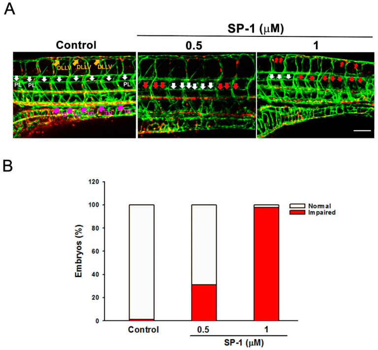
Figure 2
Heteronemin blocks thoracic duct development in zebrafish model. Transgenic (fli1:EGFP;gata1:DsRed) zebrafish embryos were treated with the indicated concentrations of heteronemin (represented as SP-1), and then thoracic duct length was analyzed via fluorescence microscopy. DLLV indicated as dorsal longitudinal lymphatic vessel (orange arrows); PL, parachordal LEC (white arrows) and TD, thoracic duct (pink arrows). (A) Representative pictures of thoracic duct of zebrafish treated with indicated concentration of SP-1 (bar = 100 μm). (B) Quantification of the defective formation of thoracic duct at 5 dpf determined by the percentages of embryos. A total of 50 embryos were analyzed in each experimental condition.