
- Title
-
Role of Extracellular Mycobacteria in Blood-Retinal Barrier Invasion in a Zebrafish Model of Ocular TB
- Authors
- Damera, S.K., Panigrahi, R.K., Mitra, S., Basu, S.
- Source
- Full text @ Pathogens

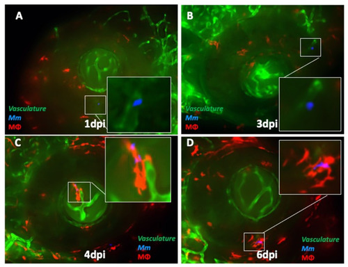
Ocular infection and granuloma formation with wild-type Mycobacterium marinum (Mm) in mpeg1:BB transgenic larvae. (A) Frequency of ocular infection following caudal vein infection with 25 colony-forming units (CFU) of Mm, 1–6 days post-injection (dpi). Granuloma formation was seen in six (33.3%) of the 18 eye-infected larvae. The kinetics of granuloma formation in all six eyes were as follows: (B) extracellular Mm (green) seeded in the eye at 1 dpi. The Mm remained extracellular until 3 dpi. (C) Mm phagocytosed inside a solitary macrophage (red) at 4 dpi. (D) Compact granuloma formation at 6 dpi, comprising macrophages with or without phagocytosed Mm. All images in the figure are from the same larva. |

Progression of ocular infection in eyes with early phagocytosis in mpeg1:BB transgenic larvae. (A) Extracellular Mycobacterium marinum (Mm, green) in the eye at 1 dpi. (B) Phagocytosis (red macrophages) is seen early at 3 dpi. (C,D) Mm remain inside solitary macrophages until 6 dpi, with no evidence of aggregation into granuloma. This sequence of events was noted in four out of 18 eye-infected larvae. All images in the figure are from the same larva. |
Kinetics of ocular infection and granuloma formation in double-transgenic (cross-bred for kdrl and mpeg1: BB) larvae with wild-type Mycobacterium marinum (Mm). (A) Extracellular Mm (blue) lying in the lumen of retinal blood vessels at 1 dpi, and (B) crossing the vascular endothelium at 3 dpi. (C) The first appearance of phagocytosis inside the retinal tissue at 4 dpi, with the infected macrophage closely abutting the vascular endothelium, and (D) aggregation of macrophages into a granuloma within the retinal tissue at 6 dpi. |
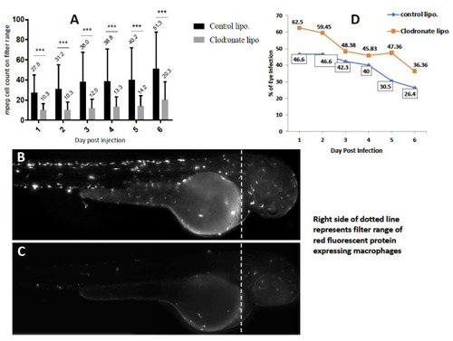
Effect of depletion of circulating monocytes on ocular infection. Liposomal clodronate was injected into mpeg1:BB transgenic larvae at 3 days post-fertilization (dpf), followed by wild-type Mycobacterium marinum injection at 4 dpf. Monocyte depletion within the head portion (shown by dashed line in B,C) was confirmed by loss of red fluorescence within the filter range at 4 dpf (Cellsense mean fluorescence intensity (MFI)) and additionally by counting manually under fluorescent microscopy. (A) MFI within the selected head area was significantly lower in the clodronate liposome-treated larvae, compared to the controls on days 1–6 post-injection (dpi) (*** p < 0.0001). (B) Representative fluorescent image (2x magnification) of control, and (C) clodronate liposome-treated larvae. (D) Higher ocular infection rate in clodronate liposome-treated larvae compared to controls on all days from 1–6 dpi. The mean percentage of larvae with eye infection in the clodronate-treated group (49.98 ± 9.57) was significantly higher than in the control group (38.73 ± 8.46) beyond 6 dpi (p < 0.03, Student’s t-test). |
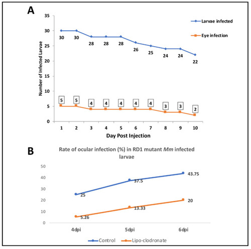
Effect of RD1 mutation in Mycobacterium marinum (Mm) on frequency of ocular infection. We infected four dpf larvae with 25 colony forming units (CFU) of RD1-mutant Mm. (A) Line graph showing significantly lower frequency of ocular infection with RD1 mutant Mm (compared with Figure 1A), even up to 10 dpi. The infected larvae, however, survived longer than during infection with wild-type (WT) Mm, as shown by the low slope of the graph. (B) Depletion of circulating monocytes with clodronate liposomes led to a drop in the ocular infection rate, the effect being opposite to that from infection with WT Mm. |
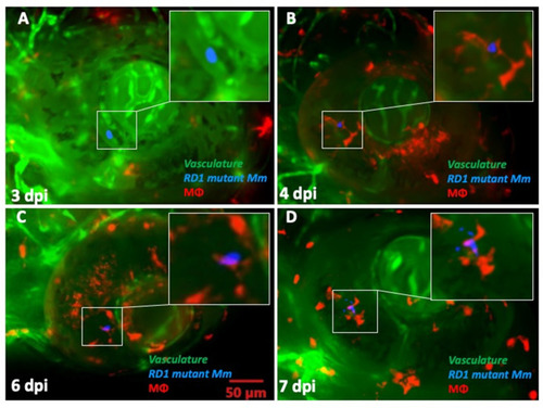
Kinetics of granuloma formation following ocular infection with RD1 mutant Mycobacterium marinum (Mm). (A) Extracellular Mm (blue) seen within retinal vascular lumen at 2 dpi. (B) Phagocytosed Mm (red macrophage) within the retinal tissue at 4 dpi. (C) Persistence of solitary macrophage with no evidence of aggregation even at 6 dpi. (D) Loose macrophage aggregation at 7 dpi, not representative of a true granuloma. This was seen in only one of the five ocular infections with RD1 mutant Mm. |
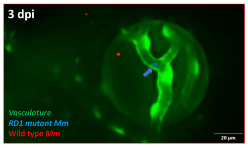
Differential progression of ocular infection by wild-type (WT) and RD1 mutant Mycobacterium marinum (Mm). At 3 dpi, WT Mm (red) have already crossed the vascular endothelial barrier and reached the retinal tissue, while RD1 mutant Mm (blue, arrow) remain inside the vascular lumen (n = 4/17). |