
IMAGE
Figure 6
- ID
- ZDB-IMAGE-210409-216
- Publication
- Damera et al., 2021 - Role of Extracellular Mycobacteria in Blood-Retinal Barrier Invasion in a Zebrafish Model of Ocular TB
- All Figures
- Figures for Damera et al., 2021
Image
|
Figure Caption
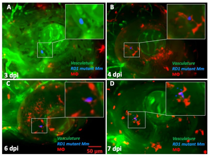
Figure 6 Kinetics of granuloma formation following ocular infection with RD1 mutant Mycobacterium marinum (Mm). (A) Extracellular Mm (blue) seen within retinal vascular lumen at 2 dpi. (B) Phagocytosed Mm (red macrophage) within the retinal tissue at 4 dpi. (C) Persistence of solitary macrophage with no evidence of aggregation even at 6 dpi. (D) Loose macrophage aggregation at 7 dpi, not representative of a true granuloma. This was seen in only one of the five ocular infections with RD1 mutant Mm.
Acknowledgments
This image is the copyrighted work of the attributed author or publisher, and
ZFIN has permission only to display this image to its users.
Additional permissions should be obtained from the applicable author or publisher of the image.
Full text @ Pathogens