
FIGURE
Figure 3
- ID
- ZDB-FIG-210409-213
- Publication
- Damera et al., 2021 - Role of Extracellular Mycobacteria in Blood-Retinal Barrier Invasion in a Zebrafish Model of Ocular TB
- Other Figures
- All Figure Page
- Back to All Figure Page
Figure 3
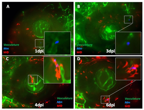
Kinetics of ocular infection and granuloma formation in double-transgenic (cross-bred for kdrl and mpeg1: BB) larvae with wild-type Mycobacterium marinum (Mm). (A) Extracellular Mm (blue) lying in the lumen of retinal blood vessels at 1 dpi, and (B) crossing the vascular endothelium at 3 dpi. (C) The first appearance of phagocytosis inside the retinal tissue at 4 dpi, with the infected macrophage closely abutting the vascular endothelium, and (D) aggregation of macrophages into a granuloma within the retinal tissue at 6 dpi. |
Expression Data
Expression Detail
Antibody Labeling
Phenotype Data
Phenotype Detail
Acknowledgments
This image is the copyrighted work of the attributed author or publisher, and
ZFIN has permission only to display this image to its users.
Additional permissions should be obtained from the applicable author or publisher of the image.
Full text @ Pathogens