
Figure 4
- ID
- ZDB-FIG-210409-214
- Publication
- Damera et al., 2021 - Role of Extracellular Mycobacteria in Blood-Retinal Barrier Invasion in a Zebrafish Model of Ocular TB
- Other Figures
- All Figure Page
- Back to All Figure Page
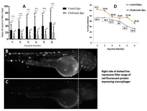
Effect of depletion of circulating monocytes on ocular infection. Liposomal clodronate was injected into mpeg1:BB transgenic larvae at 3 days post-fertilization (dpf), followed by wild-type Mycobacterium marinum injection at 4 dpf. Monocyte depletion within the head portion (shown by dashed line in B,C) was confirmed by loss of red fluorescence within the filter range at 4 dpf (Cellsense mean fluorescence intensity (MFI)) and additionally by counting manually under fluorescent microscopy. (A) MFI within the selected head area was significantly lower in the clodronate liposome-treated larvae, compared to the controls on days 1–6 post-injection (dpi) (*** p < 0.0001). (B) Representative fluorescent image (2x magnification) of control, and (C) clodronate liposome-treated larvae. (D) Higher ocular infection rate in clodronate liposome-treated larvae compared to controls on all days from 1–6 dpi. The mean percentage of larvae with eye infection in the clodronate-treated group (49.98 ± 9.57) was significantly higher than in the control group (38.73 ± 8.46) beyond 6 dpi (p < 0.03, Student’s t-test). |