
IMAGE
Figure 2
- ID
- ZDB-IMAGE-210409-60
- Publication
- Damera et al., 2021 - Role of Extracellular Mycobacteria in Blood-Retinal Barrier Invasion in a Zebrafish Model of Ocular TB
- All Figures
- Figures for Damera et al., 2021
Image
|
Figure Caption
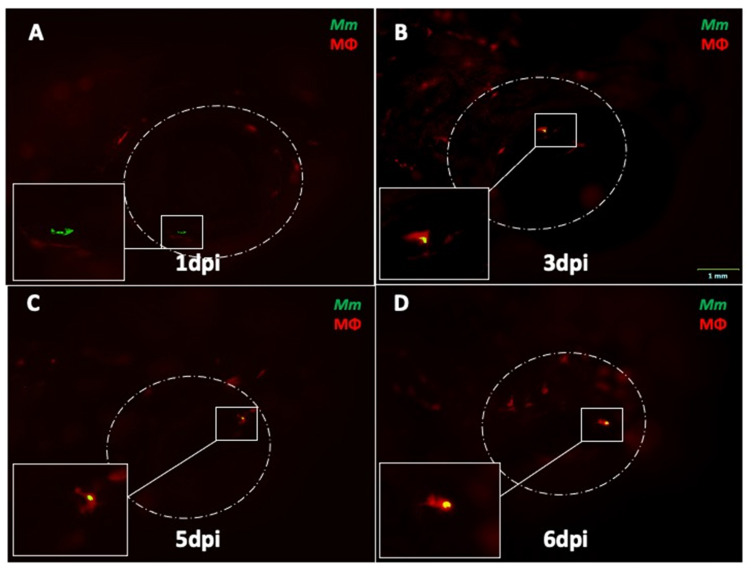
Figure 2 Progression of ocular infection in eyes with early phagocytosis in mpeg1:BB transgenic larvae. (A) Extracellular Mycobacterium marinum (Mm, green) in the eye at 1 dpi. (B) Phagocytosis (red macrophages) is seen early at 3 dpi. (C,D) Mm remain inside solitary macrophages until 6 dpi, with no evidence of aggregation into granuloma. This sequence of events was noted in four out of 18 eye-infected larvae. All images in the figure are from the same larva.
Acknowledgments
This image is the copyrighted work of the attributed author or publisher, and
ZFIN has permission only to display this image to its users.
Additional permissions should be obtained from the applicable author or publisher of the image.
Full text @ Pathogens