FIGURE SUMMARY
Title

Follistatin is a novel therapeutic target and biomarker in FLT3/ITD acute myeloid leukemia

Authors
He, B.L., Yang, N., Man, C.H., Ng, N.K., Cher, C.Y., Leung, H.C., Kan, L.L., Cheng, B.Y., Lam, S.S., Wang, M.L., Zhang, C.X., Kwok, H., Cheng, G., Sharma, R., Ma, A.C., So, C.E., Kwong, Y.L., Leung, A.Y.
Source
Full text @ EMBO Mol. Med.

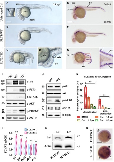
Overexpression of <styled-content toggle='no' style='fixed-case'>FLT</styled-content>3/<styled-content toggle='no' style='fixed-case'>ITD</styled-content> induced axis duplication and ectopic expression of <styled-content toggle='no' style='fixed-case'>FST</styled-content> in zebrafish embryos

The morphology of uninjected, FLT3/WT mRNA, and FLT3/ITD mRNA‐injected (150 ng per embryo) embryos on day 2 post‐fertilization (dpf).

Whole‐mount in situ hybridization (WISH) of notochord‐specific marker col9a2 in uninjected, FLT3/WT mRNA, and FLT3/ITD mRNA‐injected embryos on 2 dpf.

FLT3 signaling was detected by Western blotting in 293FT cells transfected with FLT3/ITD mRNA (I) or in zebrafish embryos injected with FLT3/ITD mRNA (J).

The effect of FLT3 inhibitor quizartinib (Qui) on the dorsalization and axis duplication phenotype induced by FLT3/ITD mRNA injection in zebrafish.

Quantification of fst expression by RT–qPCR (L), Western blotting (M), and WISH (N) after FLT3/ITD overexpression in zebrafish embryos at 6 hpf.

Data information: ov: otic vesicles; cm: cephalic mesoderm; nc: notochord. Scale bar = 500 μm. In (K and L), the experiments were performed in triplicates and the data are presented as mean ± SEM. *P < 0.05 and **P < 0.01 (Student's t‐test). NS, not significant.Source data are available online for this figure.

EXPRESSION / LABELING:
Gene:
Fish:
Anatomical Term:
Stage: Prim-5
PHENOTYPE:
Fish:
Observed In:
Stage: Prim-5

<styled-content toggle='no' style='fixed-case'>FST</styled-content> was increased in <styled-content toggle='no' style='fixed-case'>FLT</styled-content>3/<styled-content toggle='no' style='fixed-case'>ITD</styled-content>‐transgenic zebrafish and <styled-content toggle='no' style='fixed-case'>FLT</styled-content>3/<styled-content toggle='no' style='fixed-case'>ITD</styled-content>‐mutated <styled-content toggle='no' style='fixed-case'>AML</styled-content>

WISH of fst in FLT3/WT (A), and FLT3/ITD plasmid DNA‐injected zebrafish embryos without (B) or with (C) quizartinib treatment (2.5 μM) from 6 to 36 hpf. fst expression was expanded by FLT3/ITD DNA in 86% of embryos (B, arrow, 32/37) which could be effectively blocked by treating with FLT3 inhibitor quizartinib in 83% of embryos (C, 29/35).

Generation and characterization of FLT3/ITD‐transgenic zebrafish. Diagrammatic representation (D and E) of the generation of Runx1‐FLT3/ITD‐transgenic zebrafish (see Materials and Methods section). GFP expression was detected by fluorescent microscopy (F–H) and in blood circulation and thymus by WISH (I and J, blue arrow) in WT sibling and Runx1‐FLT3/ITD‐transgenic zebrafish (F1) embryos at 4 dpf. FLT3/ITD‐positive zebrafish (F1) were confirmed by PCR genotyping of GFP and FLT3/ITD using genomic DNA from fin clip of WT siblings and Runx1‐FLT3/ITD‐transgenic zebrafish (F1) at 2 months old. Fish 4, 5, and 6 showed germline transmission of FLT3/ITD transgene (K).

Kidney marrow (KM) was collected from Runx1‐FLT3/ITD‐transgenic zebrafish (F1) at 18 months old. The morphology and hematopoietic composition of KM from WT siblings (n = 6) and Runx1‐FLT3/ITD‐transgenic (n = 6) zebrafish were examined by Giemsa staining (L) and flow cytometry (M, N) (abbreviation for panel M: M, myeloid cells; P, progenitor cells; L, lymphoid cells; E, erythroid cells). Data are presented in box plot. The whiskers, boxes, and central lines in panel N represented the minimum‐to‐maximum values, 25th‐to‐75th percentile, and the 50th percentile (median), respectively. **P < 0.01 (Student's t‐test).

Expression of fst was detected by RT–qPCR in KM from WT sibling and Runx1‐FLT3/ITD‐transgenic zebrafish at 18 months old. The RT–qPCR experiments were performed in triplicates, and data were presented as mean ± SEM. **P < 0.01.

Detection of FST expression, p‐ERK1/2, and p‐CREB in mononuclear cells from normal peripheral blood stem cell (PBSC) and FLT3/ITD AML patients (diagnostic samples with leukemia blasts > 80%) by Western blotting. ^: non‐specific staining of p‐ATF1 protein due to the conserved motif.

Data information: Scale bar = 500 μm.Source data are available online for this figure.

<italic><styled-content toggle='no' style='fixed-case'>FLT</styled-content>3</italic>/<styled-content toggle='no' style='fixed-case'>ITD</styled-content> upregulated <italic><styled-content toggle='no' style='fixed-case'>FST</styled-content></italic> through phosphorylation of <styled-content toggle='no' style='fixed-case'>CREB</styled-content>

In silico analysis (DECipherment of DNA Elements, SABiosciences) and schematic model of transcription factor binding sites on human FST promoter. CBP: CREB‐binding protein; CRE: cAMP‐response element; TSS: transcription start site.

The direct binding of p‐CREB to human FST promoter was detected by ChIP‐PCR (B) and ChIP‐qPCR (C). c‐Fos was used as positive control of p‐CREB target gene. Normal IgG was used as negative control of ChIP.

Dual‐luciferase assay demonstrating the direct binding of p‐CREB on human FST promoter. pRL‐CMV, Renilla luciferase vector; pGL‐CRE− and pGL‐CRE+, firefly luciferase expression driven by human FST promoter with deleted CRE site (CRE−) or wild type (CRE+); p‐GFPSpark, GFP‐expressing vector; p‐CREBY134F, CREBY134F‐GFP‐expressing vector.

FST expression and FLT3/ITD signaling were detected by Western blotting in Ba/F3‐parental (P in short) and Ba/F3‐FLT3/ITD (ITD in short) cells.

Phospho‐flow analysis of p‐CREB in Ba/F3‐parental, Ba/F3‐FLT3/ITD, and Ba/F3‐FLT3/ITD cells treated with FLT3 inhibitor quizartinib (Qui in short). Isotype antibody was used as control to calculate the mean fluorescence intensity (MFI) ratio (F, G). The transcription and expression of Fst were detected by RT–qPCR after quizartinib treatment (10 nM) in Ba/F3‐FLT3/ITD cells for 1 day (H).

The expression of FST and phosphorylation of CREB were detected by Western blotting (I and K) and phospho‐flow analysis (J) in MOLM‐13 (I) and Ba/F3‐FLT3/ITD (K) cells treated with quizartinib and BRD7389 for 1 day, respectively.

RSK expression and FST expression were detected by Western blotting after p90RSK knockout by CRISPR/Cas9 in MOLM‐13 cells.

The phosphorylation of CREB and FST expression was detected by Western blotting in Ba/F3‐FLT3/ITD cells treated with CREB inhibitor 666‐15 for 1 day. ^: non‐specific staining of p‐ATF1 protein due to the conserved motif.

CREB expression and FST expression were detected by Western blotting after CREB knockout by CRISPR/Cas9 in MOLM‐13 cells.

The growth of Ba/F3‐parental (with IL‐3), Ba/F3‐FLT3/ITD (without IL‐3), and Ba/F3‐FLT3/ITD (with IL‐3) cells was measured after 3 days treatment of CREB inhibitor 666‐15 in vitro.

The rescue effect of CREB inhibitor 666‐15 on FLT3/ITD‐induced dorsalization and axis duplication in zebrafish embryos at 1 dpf.

Data information: In (C, D, G, H, J, and O), the experiments were performed in triplicates, and the data were presented as mean ± SEM. **P < 0.01 and ***P < 0.001 (Student's t‐test).Source data are available online for this figure.

PHENOTYPE:
Fish:
Condition:
Observed In:
Stage: Prim-5

<styled-content toggle='no' style='fixed-case'>FST</styled-content> promoted leukemia growth by activating <styled-content toggle='no' style='fixed-case'>ERK</styled-content>

FST expression in different AML cell lines was detected by Western blotting.

FST317 and FST344 overexpression resulted in significant increases in FST transcription by RT–qPCR and protein by Western blot (B) and promoted ML‐2 cell growth in vitro (C). Green, ML‐2‐GFP; blue, ML‐2‐FST317; red, ML‐2‐FST344. The RT–qPCR experiments were performed in triplicates (B).

The clonogenicity of ML‐2 overexpressing GFP, FST317, and FST344 in vitro for 14 days. The CFU experiments were performed in triplicates (E).

The engraftment of ML‐2 (with luciferase gene) overexpressing GFP, FST317, and FST344 was quantified by bioluminescence imaging (F and G), and the survival of ML‐2‐engrafted NSG mice in vivo was recorded (H). Survival curve in panel H was analyzed by log‐rank test. *P < 0.05 and **P < 0.01.

RNA‐seq and RT–qPCR validation of upregulation of RET, IL2RA, and CCL5 after FST344 overexpression in ML‐2 cells. RT–qPCR experiments were performed in triplicates (J).

Overall survival analysis of patients from TCGA‐AML based on the differential expression of IL2RA and CCL5.

Activation of MAPK/ERK pathway in ML‐2 by FST344 overexpression was validated by Western blotting. Scale bar = 1 mm.

Data information: In (B, C, E, G, and J), data were presented as mean ± SEM. *P < 0.05, **P < 0.01, and ***P < 0.001 (Student's t‐test). ns: not significant.Source data are available online for this figure.

<italic><styled-content toggle='no' style='fixed-case'>FST</styled-content></italic> knockdown reduced <styled-content toggle='no' style='fixed-case'>AML</styled-content> cell growth <italic>in vitro</italic> and <italic>in vivo</italic>

FST knockdown in MOLM‐13 by shRNA effectively reduced FST expression.

The morphology (B), apoptosis (C), and clonogenicity of MOLM‐13 (D and E) were measured after FST knockdown in vitro. Scale bar = 10 μm. The apoptosis assays (C) were performed in triplicates.

The engraftment of MOLM‐13 after FST knockdown was detected by flow cytometry of human CD45‐ and mouse CD45.1‐positive cells in recipient mouse BM aspiration at week 2 post‐transplantation.

The effect of FST knockdown on the survival of NSG mice engrafted with MOLM‐13. scr: scrambled sequence control (7 mice); sh: short hairpin RNA (8 mice for sh1 and sh2, respectively). The survival curve was analyzed by log‐rank test. **P < 0.01.

Data information: In (C, E, and G), data were presented as mean ± SEM. *P < 0.05 and **P < 0.01 (Student's t‐test).Source data are available online for this figure.

<italic><styled-content toggle='no' style='fixed-case'>FST</styled-content></italic> targeting by <styled-content toggle='no' style='fixed-case'>CRISPR</styled-content>/Cas9 or antisense oligo significantly reduced leukemia cell growth <italic>in vitro</italic> and <italic>in vivo</italic>

The morphology and clonogenicity of MOLM‐13 after FST knockout by CRISPR/Cas9 in vitro. Scale bar = 10 μm.

The engraftment of MOLM‐13 after FST knockout was detected by flow cytometry of human CD45‐ and mouse CD45.1‐positive cells in recipient mouse BM aspiration at week 2 post‐transplantation.

The effect of FST knockout on the survival of NSG mice engrafted with MOLM‐13 cells. Cas9: Cas9 only (10 mice); sgRNA#3: Cas9 + sgRNA#3 (10 mice); sgRNA#4: Cas9 + sgRNA#4 (8 mice).

The knockdown efficiency of different FST‐specific antisense oligos (ASOs) in MOLM‐13 cells was detected by RT–qPCR after 3 days of treatment in vitro. The knockdown and RT–qPCR experiments were performed in triplicates.

MOLM‐13 cell growth was measured after 3 days of treatment of FST‐ASO in vitro. The ASO treatment experiments were performed in triplicates.

Intraperitoneal injection of FST‐ASO (10 mg/kg weekly, 6 mice) significantly prolonged the survival of MOLM‐13‐engrafted NSG mice. The random sequence was used for negative control (Neg‐ASO, 6 mice).

Data information: In (B, D, F, and G), data were presented as mean ± SEM. *P < 0.05 and **P < 0.01 (Student's t‐test). In (E and H), survival curves were analyzed by log‐rank test. *P < 0.05.

Serum <styled-content toggle='no' style='fixed-case'>FST</styled-content> correlated with leukemia progression and therapeutic response in mouse and human <italic><styled-content toggle='no' style='fixed-case'>FLT</styled-content>3</italic>/<styled-content toggle='no' style='fixed-case'>ITD AML</styled-content>

Flt3/ITD knock‐in mouse were genotyped (A). The spleen weight (B and C, 4 mice each) and serum Fst level (D) were measured in WT siblings and Flt3/ITD knock‐in mice (8 mice each). In (C and D), data were presented in box plot. The whiskers, boxes, and central lines represented the minimum‐to‐maximum values, 25th‐to‐75th percentile, and the 50th percentile (median), respectively. **P < 0.01 (Student's t‐test).

The engraftment of MOLM‐13 in NSG mice was confirmed at week 1 post‐injection (E, F). Serum FST level was measured in MOLM‐13‐engrafted NSG mice at pre‐injection and week 2 post‐injection (G, PBS group, 3 mice; MOLM‐13 group, 4 mice). After FST knockdown, serum FST level was also measured in MOLM‐13‐engrafted NSG mice at week 2 post‐injection (H, 3 mice for each group).

Serum FST level was significantly increased in primary AML‐derived xenografted mouse at week 6 post‐injection. Human primary AML cells (FLT3/ITD‐positive, leukemia blasts > 80%, 10 × 106, n = 7) were injected via tail vein into irradiated NSG mice at 6–8 weeks old (I). Human leukemic engraftments were confirmed by flow cytometry of human CD45 and mouse CD45.1 cells in recipient mouse BM aspiration (J). Serum FST from pre‐injection and post‐engraftment mouse was measured (K, one mouse for each primary AML sample).

Correlation between serum FST levels and leukemia blast percentage from FLT3/ITD‐mutated AML at diagnosis. Correlation analysis (Pearson's correlation coefficient) was performed by GraphPad Prism 6.

Serum FST decreased in CR and increased after relapse in 4 AML patients receiving quizartinib monotherapy.

Serum FST continued to rise during disease progression from a patient who did not respond to quizartinib. Patients in (M) and (N) were recruited in the QUANTUM‐R, and patient accrual has been completed.

Data information: In (G and K), data were presented as scatter dot plot. n.s: not significant, *P < 0.05 and **P < 0.01 (Student's t‐test). In (H, M and N), data were presented as mean ± SEM. *P < 0.05 and **P < 0.01 (Student's t‐test).

Schematic diagram depicting the molecular mechanism of <styled-content toggle='no' style='fixed-case'>FST</styled-content> upregulation in <styled-content toggle='no' style='fixed-case'>FLT</styled-content>3/<styled-content toggle='no' style='fixed-case'>ITD</styled-content>‐transgenic zebrafish, knock‐in mice, and human <styled-content toggle='no' style='fixed-case'>AML</styled-content>

Left panel. Transient overexpression of human FLT3/ITD mutation resulted in axis duplication and dorsalization abnormalities in zebrafish accompanied by upregulation of embryonic morphogen Fst. Upregulation of FST was consistently found in FLT3/ITD‐transgenic zebrafish, Flt3/ITD knock‐in mice, FLT3/ITD‐mutated AML cell lines, and primary AML samples in vitro and in vivo. Right panel. A novel FLT3/ITD‐p90RSK‐CREB‐FST signaling cascade was demonstrated in human AML. FST is a promising biomarker and therapeutic target for human FLT3/ITD+ AML.

Acknowledgments
This image is the copyrighted work of the attributed author or publisher, and ZFIN has permission only to display this image to its users. Additional permissions should be obtained from the applicable author or publisher of the image. Full text @ EMBO Mol. Med.