FIGURE SUMMARY
Title

MicroRNA26 attenuates vascular smooth muscle maturation via endothelial BMP signalling

Authors
Watterston, C., Zeng, L., Onabadejo, A., Childs, S.J.
Source
Full text @ PLoS Genet.

<italic>miR26a</italic> is expressed in blood vessels; endothelial cells have active BMP signalling.

A) Model of how miR26a controls BMP signaling via direct targeting of smad1. B) Lateral view of whole mount in situ expression of miR26a at 48 hpf shows ubiquitous expression pattern, with strong expression in the ventral head of the embryo. B’) Cross section of the head at 48 hpf. C) At 4 dpf miR26a is expressed in the pharyngeal arches, bulbous arteriosus and ventral aorta. C’) Cross section of the head showing miR26a expression in blood vessels (purple; punctate stain) compared with endothelial stain (brown; kdrl:GFP transgenic). Inset is an enlargement of image in C’. D) Ventral view of the pharyngeal region of a 4 dpf double transgenic Tg(BRE:EGFP);Tg(kdrl:mCherry) embryo shows BRE:EGFP (green) expression within endothelial cells in aortic arches (red, white arrowheads in E’-E”‘) and ventral aorta (red, white arrowheads F’-F”‘). G-H) Ventral and lateral views of a 4 dpf double transgenic Tg(BRE:EGFP); Tg(acta2:mCherry) zebrafish shows that acta2 positive cells are in direct contact with BMP-responsive endothelial cells but do not express BRE:EGFP. Scale bar represents 50μm.

<italic>miR26a</italic> knockdown increases <italic>smad1</italic> expression.

A) Schematic of miR26a transient knockdown methods. B and C) Relative expression level of miR26a in morpholino and CRISPRi injected embryos at 48 hpf (n = 3). D) Whole mount in situ hybridization staining for smad1 at 48 hpf and 4 dpf shows increased expression of smad1 in miR26a knockdown embryos particularly in the ventral aorta, aortic arches and pharyngeal region (dotted outline). Scale bar represents 200μm. E) Relative expression of smad1 in 48 hpf morphants is increased compared to control embryos (n = 3). F) Relative expression of smad1 in 4 dpf miR26a morphants is increased compared to control embryos (n = 4). RT-qPCR data show the mean ± SEM, Student's two-tailed t-test *p < 0.05, n = number of biological replicates.

<italic>miR26a</italic> knockdown embryos have increased endothelial pSmad1.

A-D) Ventral view confocal projections of the 4 dpf ventral aorta (dotted outline). Endothelial nuclei (fli1a:nEGFP; A-D, arrowheads) and pSmad1/5/9 (pSmad1,white A’- D’) and overlay (magenta, A”- D”) in 4 dpf Scr. Control (A), miR26a MO (B), dCas9 control (C) and miR26a CRISPRi (D) embryos. Yellow arrowheads indicate double positive pSmad1 + fli1a:nEGFP nuclei in the ventral aorta. E) Quantification of total number of fli1a:nEGFP nuclei in the ventral aorta. F-G) Quantification of the percentage of double pSmad1; fli1a:nEGFP positive nuclei in miR26a morphants (F) and miR26a CRISPRi (G) embryos. N = 3 experiments. Total embryos are as follows: Scr. Control MO n = 14, miR26a MO n = 14, dCas9 control n = 9 and miR26a CRISPRi n = 10. Student's two-tailed t-test, p***< 0.0001 and p ****< 0.00001 as compared to WT, error bars = SD. Scale bar: represents 50μm.

Increased levels of <italic>smad1</italic> result in defects in the vascular system and body axis.

A-C) Representative 48 hpf miR26a knockdown embryos with hemorrhage, as indicated by arrows. D) Quantification of average rates of hemorrhage. (Error bars = SD Unpaired t test, miR26a MO *p< 0.01 and mi26 CRISPRi **p< 0.001 as compared to WT, N = 3, Wildtype n = 224, miR26a MO n = 124, miR26a CRISPRi n = 180). E-F) Representative morphology after smad1 overexpression. G-I) miR26a and smad1 double knockdown experiments. G) Representative 48 hpf smad1 MO embryos with mild (V1) and severe (V2) ventralization phenotypes. H) Representative 48 hpf double miR26a and smad1 knockdown embryos with rescued hemorrhage and normal body axis showing only mild (V1-WT) ventralization phenotypes. I) Quantification of observed phenotypes double knockdown experiments (N = 4, total n wildtype = 193, Scr. Control MO = 157, smad1 MO = 175, smad1 mRNA = 95, miR26a MO = 190, and miR26a MO + smad1 MO = 190. One Way ANOVA of hemorrhage phenotype; Wildtype/Scr. Control MO vs. miR26a MO p< 0.0001 Wildtype/Scr. Control vs. SMAD1 mRNA p< 0.0001 miR26a MO vs. miR26a MO+ smad1 MO p< 0.0001 One Way ANOVA of V1 phenotype: Wildtype/Scr. Control MO; vs. smad1 MO p< 0.0001 vs. miR26a MO+ smad1 MO p< 0.0001. Error Bars = SEM. Scale bar represents 500μm.

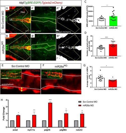
Loss of <italic>miR26a</italic> morphants leads to increased expression of vSMC genes and <italic>acta2</italic>-positive vSMCs.

A-B) Representative ventral views of 4 dpf Tg(BRE:EGFP); Tg(acta2:mCherry) embryos. Scr. Control embryos (A-A”) and miR26a morphant embryos (B-B”) showing qualitative upregulation of BRE:EGFP in the ventral aorta (VA) and pharyngeal arch arteries (PAA). C) Quantification of green fluorescent marker (BRE:EGFP) along the VA, taken from the highlighted yellow region in A’ and B’, and represented as corrected total cell fluorescence (CTCF) (N = 3, miR26a MO n = 15, Scr. Control n = 12, Unpaired t test, ****p< 0.0001 as compared to control, error bars = SEM). D) Quantification of acta2 positive cell number on VA and PAAs, within area outlined in A” and B”. Number of acta2 positive cells is significantly increased in miR26a morphants (N = 3, miR26a MO n = 18, Scr. Control n = 15, Unpaired t test, ****p< 0.0001 as compared to control, error bars = SEM). E and F) Measurement of vSMC height (yellow axis) from the endothelium (white dashed line). Representative images of ventral aorta (from insets), Scr. Control (E) and miR26a morphants (F). G) Quantification of average vessel heights along the length of the VA (N = 3, miR26a MO n = 18, Scr. Control n = 13, Student's two-tailed t-test, ****p< 0.0001 as compared to control, error bars = SEM). H) RT-qPCR quantification of vSMC differentiation genes in injected controls and miR26a morphants (n = 3). RT-qPCR data show the mean ± SEM, Student's two-tailed t-test *p < 0.05, n, number of biological replicates.

<italic>smad1</italic> overexpression in endothelial cells results in increased vSMC coverage.

A) Vector construct for overexpression of smad1 under the endothelial cell promoter kdrla. B) RT-qPCR fold change in smad1 and id-1 expression levels in endothelial specific smad1 overexpressing embryos (smad1ECOE) embryos at 4 dpf (n = 3). RT-qPCR data show the mean ± SEM, Student's two-tailed t-test *p < 0.05, n, number of biological replicates. C–D) Representative orthogonal projections of ventral views of 4 dpf Tg(BRE:EGFP); Tg(acta2:mCherry) embryos. Control embryos (C-C”) and smad1ECOE embryos (D-D”) showing endothelial BRE:EGFP and vSMC acta2:mCherry expression in the ventral aorta (VA) and pharyngeal arch arteries (PAA). E) Quantification of green fluorescent marker (BRE:EGFP) along the VA, highlighted within the yellow region in C” and D”, as corrected total cell fluorescence (CTCF). F) Quantification of acta2 positive cell number on VA, within area outlined in C and D. Number of acta2 positive cells is significantly increased in smad1ECOE embryos. G) Quantification of length of VA, within area outlined in C and D. H) Quantification of the percent vSMC coverage of ventral aorta. For each quantification, N = 3, smad1ECOE embryos n = 8, Control n = 8, Student's two-tailed t-test, *-***p< 0.01–0.0001 as compared to control. Error bars = SEM, Scale bar represents 50μm.

<italic>miR26a</italic> controls vSMC differentiation via <italic>smad1</italic>-mediated BMP signaling.

Ventral aorta showing endothelial (red) and smooth muscle (green) cells in miR26a morphants or CRISPRi- injected embryos treated with vehicle control (DMSO) or 15μM K02288 from 52 hpf to 4 dpf. A-C) DMSO-treated vehicle control embryos. D-F) K02288 treated control embryos. (A, D), miR26a morphant (B, E), miR26a CRISPRi knockdown (C, F). A’-F’ are threshold adjusted images of acta2-EGFP expression. G) Quantification of acta2 positive cell number on VA and PAAs, within area outlined in A and B. Number of acta2 positive cells is significantly reduced in K02288 treated embryos as compared to DMSO control. There is no significant decrease in miR26a knockdown embryos (two Way ANOVA, N = 3, miR26a MO n = 15, Wildtype n = 15, Unpaired t test, ****p< 0.0001 as compared to control, Error Bars = SEM. H) Quantification of length of VA, within area outlined in A and B. Length of VA is significantly reduced in K02288 treated embryos as compared to DMSO control. There is no significant decrease in miR26a knockdown embryos (Two Way ANOVA, N = 3, miR26a MO n = 15, Scr.Control n = 15, Unpaired t test, ****p< 0.0001 as compared to control, Error Bars = SEM. VA = ventral aorta, HA = hyoid artery, BA = bulbous arteriosus. (N = 3, 8–9 embryos per treatment group. One Way ANOVA, p< 0.001–0.0001***-****. Scale bar represents 50μm. I-L) pSmad1/59 staining in K02288 treated embryos. Endothelial nuclei (fli1a:nEGFP; I-L, arrowheads) and pSmad1/5/9 (pSmad1, white I’-L’) and overlay (magenta, I”-L”) in 4 dpf Scr. Control and miR26a morphants. Solid yellow arrowheads in I”-L” indicate pSmad1 + fli1a:nEGFP double positive nuclei in the ventral aorta. M) Quantification of total number of fli1a:nEGFP nuclei in the ventral aorta. N) Quantification of the percent pSmad1; fli1a:nEGFP double positive nuclei in miR26a morphants and miR26a CRISPRi embryos. N = 3 experiments, total embryos Scr. Control MO n = 9, miR26a MO n = 9. One Way ANOVA, p< 0.001–0.0001***-****. Scale bar represents 50μm.

Mechanistic model by which <italic>miR26a</italic> modulates BMP signaling to promote vSMC differentiation via interactions with endothelial cells.

miR26a modulates vascular stability by directly targeting smad1. At developmental stages when smooth muscle appears, the endothelium has active BMP signaling. Loss of miR26a results in increased BMP signaling in endothelial cells where Smad1 becomes phosphorylated. Increased pSmad1 in endothelial cells leads to increased differentiation (acta2 expression) and increased vSMC cell number, while blocking BMP signaling leads to a decrease of both. (Dashed arrows indicated the indirect effect on vSMC marker expression and cell number).

ZFIN is incorporating published figure images and captions as part of an ongoing project. Figures from some publications have not yet been curated, or are not available for display because of copyright restrictions.

ZFIN is incorporating published figure images and captions as part of an ongoing project. Figures from some publications have not yet been curated, or are not available for display because of copyright restrictions.

EXPRESSION / LABELING:
Gene:
Fish:
Knockdown Reagent:
Anatomical Term:
Stage: Long-pec
PHENOTYPE:
Fish:
Knockdown Reagent:
Observed In:
Stage: Long-pec

ZFIN is incorporating published figure images and captions as part of an ongoing project. Figures from some publications have not yet been curated, or are not available for display because of copyright restrictions.

EXPRESSION / LABELING:
Genes:
Fish:
Knockdown Reagent:
Anatomical Term:
Stage: Day 4
PHENOTYPE:
Fish:
Knockdown Reagent:
Observed In:
Stage: Day 4
Acknowledgments
This image is the copyrighted work of the attributed author or publisher, and ZFIN has permission only to display this image to its users. Additional permissions should be obtained from the applicable author or publisher of the image. Full text @ PLoS Genet.